
Utilizing a digital workflow for cosmetic crown lengthening, an economical and efficient must-have tool
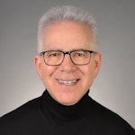
After seeing the great return on investment his brother received from Dr. Glassman’s New York City dental office, one patient seized the opportunity for a consultation of his own. As a healthy 22-year-old college student, the patient sought out dentistry to enhance his facial esthetics and offer his smile a well-deserved promotion (figure 1).

During the initial appointment, the patient stated that his objective was to lessen the amount of gums he displayed while smiling. Records were obtained, including facial photos, intraoral scan, and CBCT. Through quantitative analysis, the height-to-width ratio of his anterior central teeth was nearly 1:1, with the ideal ratio of width to height being 0.80. Also noted was the wear on the teeth. This caused the teeth to be 8 mm in length when they should be between 10 mm and 11 mm.
A series of diagnoses were made, which included excess vertical maxilla growth, altered active eruption, and hypermobility of the upper lip. The treatment plan included two parts: a “smile lift” by repositioning the maxillary alveolar crest apically and minimally invasive porcelain veneers on teeth nos. 4–13. This would add an additional 3 mm to the overall appearance of the central incisors as well as lengthening the additional teeth included in the treatment plan.
What was unique in this case was that it was designed digitally using facially driven design principles. Patients want to see the desired outcome before making an investment and undergoing dental procedures.
Traditional analog process
First, let’s review the traditional analog way of doing this. The traditional approach to an osseous crown lengthening procedure entails a discussion between the restorative dentist and the periodontist based on the photos. Often, in the photos and/or on a separate model, the new tissue height would be drawn and material added to the incisal edge would be marked on the photo or “waxed up” manually on a model. In this case, roughly 2 mm of gingival tissue would be removed, followed by adding 1–2 mm in incisal length.
The periodontist would perform the procedure by anesthetizing the patient to perform bone sounding to determine the distance from the most coronal position of alveolar bone to the cementoenamel junction (CEJ) of the patient’s original tooth position.
Typically, after bone sounding, the periodontist will mark on the gingiva with indelible ink, reflect a full thickness flap and perform the osseous surgery. Biological width is maintained by reflecting a full-thickness tissue flap and electing to reduce the bone in an apical direction (3–4 mm apical to the newly selected most apical portion of the crown) to show the desired amount of tooth after the tissue is excised, while maintaining a minimum of 2 mm of keratinized tissue around the new gingival margin. The flap is then approximated at the new and ideal position on the teeth and sutured closed for healing purposes. Approximately 0.5 mm apical gingival creep is anticipated, as it will ultimately follow the height and contour of the underlying bone.
After eight weeks of healing, the patient would be referred back to the restorative dentist, where new impressions would be taken, new photos taken, and a new wax-up made that would be used as a reduction guide and a matrix to create the provisionals.
Digital approach to osseous crown lengthening
The new digital approach (figures 2–13) to an osseous crown lengthening procedure involves photographs, radiographs, intraoral scans, and CBCT data for a facially driven treatment plan. This approach starts with the end result in mind and ultimately requires fewer appointments for the patient. It also leads to more predictable esthetic results.


The digital approach allows the patient to visually see the new length of their teeth relative to the lip when smiling and understand the process of the proposed treatments. The doctor can explain in detail how each procedural step helps alleviate the patient’s chief complaint of a gummy smile. The predictability of the newly accepted treatment plan is additionally enhanced by having the specialists and the lab on the same page regarding the end goal from the beginning of treatment, rather than typically being involved toward the end of the journey.
Once the patient accepts treatment and is on board for the crown lengthening as proposed, a surgical guide stent must be created and printed for approximation of the new crown margins. Using the smile creator Exocad, a digital mock-up of the final margins of the newly lengthened teeth is generated. The patient can be presented with the simulated smile and accept the look of the mock-up before digitally fabricating the surgical guide needed to achieve said results.
Prior to treatment, the patient mentioned he had previously undergone orthodontic treatment. After analyzing all intraoral iTero scans and an occlusogram to account for restorative functionality, the patient was found to have a class 1 sagittal relationship, adequate arch length within the transverse plane, and normal 33% overbite across the vertical plane. His occlusal relationships in each dimension were verified to be functional and healthy, meaning that no additional alignment adjustments were necessary.
Utilizing a facially driven treatment plan, the facial photographs were merged with the CBCT scan to assess bone levels, and the central incisors were digitally planned to exhibit a new length of 11.5 mm, to enhance the patient’s long facial structure. The new width of the veneers could then be generated to be 85% of this measurement to achieve the golden proportion for optimal esthetics. Once the surgical guide is designed in Exocad to achieve these coronal margins, the guide design is exported to SprintRay to be printed with a snug fit for accurate coronal margination for the final restorations later on.
New scans of the patient’s mouth were taken eight weeks after the osseous surgery, prior to the minimal veneer preparations. The original digital plan then can be evaluated, which typically involves minor changes. The next phase design would be printed in the office following sharing the STL files (stereolithography). This allows the office to create a restorative matrix and reduction guide for the dentist. Careful preparation was utilized so as to stay within enamel only, which many studies have shown allows for more predictable bonding and longevity of veneer restorations. Standard tissue retraction methods were used, and the preps were scanned with iTero 5D Plus. The provisionals created from the digital plan would allow the patient to test-drive the new smile prior to the final 10 porcelain restorations being placed. The patient was seen a few days after his preps to allow any modifications before the final 10 e.max pressable veneers were created.
After experiencing the comparative advantage the provisional teeth offered him, the patient was ready for the final restorations to be placed. After placement, the patient left the office with both an esthetic and functional upgrade to take on life after college with confidence, featuring 10 porcelain veneers across his maxillary teeth.
As a review, the digital approach greatly enhanced an outcome built upon accurate and predictable surgery, conservative tooth preparation, and tailored esthetics that both enhanced the patient’s natural features and addressed his main concern in an efficient and well-communicated series of steps.
Editor's note: This article appeared in the February 2025 print edition of Dental Economics magazine. Dentists in North America are eligible for a complimentary print subscription. Sign up here.


, a collaborative approach to dentistry where oral health is integrated into the entirety of patient care")
