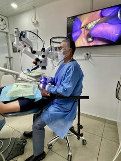
Factors constraining the widespread integration of dental microscopy in clinical practice
Integration of the dental microscope into clinical practice remains a topic of contention within the dental community, often encountering resistance that merits scholarly exploration. As a seasoned practitioner and educator with extensive experience in utilizing dental microscopy, I have observed and analyzed the reasons underlying this reluctance. This review delves into the multifaceted barriers that impede the widespread adoption or appropriate utilization of dental microscopy in clinical settings.
The dental microscope represents a pinnacle in precision dentistry, yet its acceptance within the dental profession is not universal. While many specialists, particularly endodontists,1 have seamlessly integrated this technology into their practice, a substantial portion of general dentists remain hesitant.2 I believe this hesitancy warrants investigation.
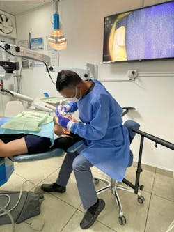
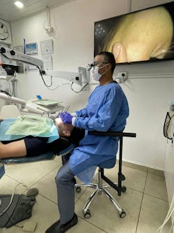
Conventional naked-eye or loupes practice has been shown to have limitations in offering the precise visual information we need to reach quality outcomes for complex dental procedures.3 This practice also deteriorates dentists’ lives outside of the office due to unbearable pain in the neck, back, shoulder, or wrists, even putting their professional practice at risk (figure 1).4
There are some general misconceptions within the dental community regarding the use of the dental microscope. Some believe:
- The microscope is only practical for endodontics.
- Dentists can achieve the necessary magnification for precision work with their magnifying glasses or even with the naked eye.
- Microscopes are too expensive, and there is no return on investment.
However, some hidden factors prevent dentists from considering incorporating this precision tool.
Educational exposure
A pivotal determinant in shaping professional practices is the exposure received during the formative years of dental education. Habits and methodologies acquired during training often persist into professional careers. Unfortunately, the curricula of many dental schools inadequately emphasize the ergonomic benefits and procedural enhancements facilitated by dental microscopy.4 This deficiency in exposure diminishes awareness of its potential advantages, including improved posture and enhanced visual acuity. Early integration of microscopy into dental education is essential to instill proper ergonomic principles and foster appreciation for its utility.
Learning curve and comfort zone
One of the biggest challenges for humans is facing their comfort zone. Transitioning to a novel tool such as the dental microscope necessitates overcoming the inertia of established practices and venturing beyond one’s comfort zone. Dentists accustomed to conventional methods may harbor apprehensions regarding the learning curve associated with microscopy. Mastery of this technology requires perseverance, guided repetition, and adaptation to new working methodologies.5,6 Central to this process is recognizing that proper utilization of the microscope engenders ergonomic benefits, mitigating physical strain and optimizing procedural efficiency. Encouraging dentists to embrace this paradigm shift is imperative to realizing the transformative potential of dental microscopy.7
Economic considerations
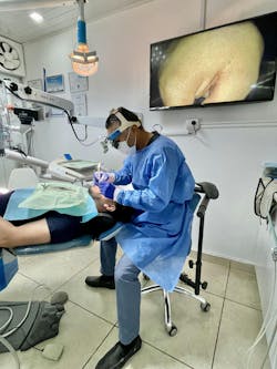
The cost of dental microscopes ranges from $20,000 to $50,000. This presents a substantial investment for dental practices, particularly in light of other operational expenses. However, juxtaposed against the staggering prevalence of musculoskeletal disorders (MSDs) and early retirement or disabilities due to workplace MSDs, this investment assumes a dual significance for dental professionals.8,9 Proactive investment in ergonomic solutions, such as dental microscopy, not only safeguards the well-being of the practitioner but also augments treatment quality and patient satisfaction. Incorporating video systems within microscopes enhances patient engagement and fosters a favorable ROI through enhanced diagnostic elucidation and treatment communication.10
The dental microscope elevates the standard of care
The integration of dental microscopy into clinical practice demands a multifaceted approach that addresses educational, psychological, and economic considerations. Early exposure within dental education, coupled with targeted interventions to alleviate apprehensions and incentivize investment, holds promise for fostering widespread adoption of this tool. Embracing dental microscopy transcends disciplinary boundaries, offering tangible benefits to practitioners and patients alike. By nurturing a culture of innovation and ergonomic consciousness, the dental profession can elevate the standard of care while safeguarding the well-being of its clinicians.
Editor's note: This article originally appeared in DE Weekend, the newsletter that will elevate your Sunday mornings with practical and innovative practice management and clinical content from experts across the field. Subscribe here.
References
- Setzer FC. Endodontics Colleagues for Excellence: The dental operating microscope in endodontics. American Association of Endodontics. Winter 2016. https://www.aae.org/specialty/wp-content/uploads/sites/2/2017/07/winter2016microscopes.pdf
- Gupta A, Bhat M, Mohammed T, Bansal N, Gupta G. Ergonomics in dentistry. Int J Clin Pediatr Dent. 2014;7(1):30-34. doi:10.5005/jp-journals-10005-1229
- Atlas AM, Janyavula S, Elsabee R, et al. Comparison of loupes versus microscope-enhanced CAD-CAM crown preparations: a microcomputed tomography analysis of marginal gaps. J Prosthet Dent. 2024;131(4):643-651. doi:10.1016/j.prosdent.2022.04.008
- Pispero A, Marcon M, Ghezzi C, et al. Posture assessment in dentistry for different visual aids using 2D markers. Sensors (Basel). 2021;21(22):7717. doi:10.3390/s21227717
- Friedman M, Mora AF, Schmidt R. Microscope-assisted precision dentistry. Compend Contin Educ Dent. 1999;20(8):723-728,730-731,735-736;quiz 737.
- Bud M. Pricope R, Pop RC, et al. Comparative analysis of preclinical dental students’ working postures using dental loupes and dental operating microscope. Eur J Dent Educ. 2021;25(3):516-523. doi:10.1111/eje.12627
- Steidley A, Kersten D, Delgado S, Mines P, Beltran T. Use of the microscope in endodontics: a questionnaire-based study [abstract]. In: J Endod. 2018;44(3):Abstract PR50.
- Duckworth AL, Gross JJ. Behavior change. Organ Behav Hum Decis Process. 2020;161(Suppl):39-49. doi:10.1016/j.obhdp.2020.09.002
- Hendrick HW. The ergonomics of economics is the economics of ergonomics. Proceedings of the Human Factors and Ergonomics Society Annual Meeting. 1996;40(1):1-10. doi:10.1177/154193129604000101
- Clark D. A microscope for every dentist: why and how. Dentaltown. January 2010. https://www.dentaltown.com/magazine/article/2467/a-microscope-for-every-dentist-why-and-how
Juan Carlos Ortiz Hugues, DDS, CEAS, is a master of the Academy of Microscope Enhanced Dentistry (AMED), associate professional in ergonomics by BCPE, CEAS II, and president of the AMED. He wrote the book, Ergonomics Applied to Dental Practice, available from Quintessence Publishing. He provides lectures, training, and advice on advanced dental ergonomics in Latin America and the United States, mainly related to ergonomics applied to dental microscopy.

