Robert A. Lowe, DDS, FACD, FADI, FAGD, FASDA, FIADE
Placing tooth-colored direct composite restorations is a clinical challenge even for the most seasoned clinician. Aside from the technique sensitivity involved in adhesive bonding, finding a shade—whether single or layered—that predictably and precisely matches the natural tooth consistently can be extremely difficult. Shade taking for most composite materials relies on 1950s technology, the Vita Lumin shade guide. The caveat is that for many brands, A1 composites don’t precisely match the Vita A1 shade guide, let alone one another! This can be extremely frustrating for the clinician and especially for patients who are particular about esthetic matching. It is even harder when using “bleach shades,” as there is little to no standardization for those high-value materials.
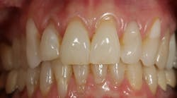
Figure 1: Preoperative retracted facial view showing tooth No. 7, which is considerably shorter than No. 10 on the contralateral side. This is an esthetic concern for the patient.
Having a universal, single shade of composite material that takes the guesswork out of shade selection and matches the majority of clinical situations is probably on most clinicians’ wish lists. The following is a case report that describes the use of a new composite material, Omnichroma (Tokuyama Dental America), which just may fill that niche.
Figure 2: A Vita Shade tab (B1) is displayed as a reference to the original tooth shade that is being matched in the restoration.
Omnichroma is the first “omnichromatic” composite material where one shade of composite will virtually match any shade on any patient. Natural tooth color typically falls in the red to yellow area of the color spectrum. Most composite materials depend on the chemical color of the resin to fall in that area of the spectrum and to match tooth color. This means that an A1 composite is chemically colored to specifically match an A1 tooth. For this reason, multiple shades are needed to cover all patients and all clinical situations. Omnichroma’s proprietary technology utilizes structural color rather than chemical coloration as its main color mechanism. As light passes through the composite resin’s spherical fillers, the light is altered along the red and yellow areas of the color spectrum to take on and match the color of the patient’s tooth.
Figure 3: A 3–4 mm bevel is placed using a pointed diamond bur (Komet USA) from the mesiofacial line angle to the distofacial line angle with the horizontal component being curved so the restorative material will blend seamlessly with the surrounding tooth color.
Clinical case report
This 52-year-old female patient, whose retracted smile view is shown in Figure 1, reported with a chief complaint that her “right front tooth (she pointed to her right maxillary lateral incisor) was shorter than the same tooth on the opposite side” (left maxillary lateral incisor). Her dental history included generalized attachment loss (gingival recession) with concomitant bone loss. Several teeth had Class V composites (not good esthetic matches!) placed previously due to root surface decay secondary to gingival recession and root surface exposure. Her occlusion was evaluated in all excursions to see if she could accommodate additional length being added to tooth No. 7 to satisfy her chief complaint, while her case was worked up to address other potential long-term solutions.
Figure 4: Universal Bond (Tokuyama Dental America) is mixed in a disposable dappen dish.
The preoperative shade appears to be close to a Vita Lumin B1 shade (figure 2). As previously discussed, matching a direct composite material to tooth structure is a clinical challenge in itself, let alone trying to match a portion of a single tooth seamlessly with the surrounding enamel, given internal characterization and the gradation of opacity and translucency in the local area where the composite material overlaps the natural tooth surface. One unique feature of Omnichroma is that it takes away the dentist’s worry of the restored tooth not exactly matching the shade guide, as well as the unpredictability of choosing the correct shade or having to mix multiple shades in an attempt to match the patient’s tooth.
Figure 5: The bonding agent is applied to the cavity preparation.
It is also important to create a long bevel of enamel (2–3 mm) with about 0.5–0.75 mm of actual tooth reduction facially to allow the restorative material to have sufficient thickness to give it a chance to blend with the natural tooth (figure 3). This is critical so that there is enough enamel to bond sufficiently and retain the restoration. A common mistake is not to reduce (prepare) enough depth to give the composite material sufficient thickness to optically blend with the surrounding tooth or create enough surface area with which to bond. The proximal margins of the preparation extend to the proximal-facial line angles respectively. This is because it is easier to blend restorative material with tooth structure if the margin is located at the intersection of two geometric surfaces of the tooth rather than in the middle of the facial surface. In this case, there is no need to involve the proximal contact areas in the preparation. The horizontal margin located at the junction of the incisal and middle third of the tooth is curved mesial to distal to make the restorative material margin less visible after finishing.
Figure 6: Omnichroma Blocker (Tokuyama Dental America) is placed to build a lingual shelf to the preparation extending the cervicoincisal length to more closely match the contralateral maxillary lateral incisor.
Once the preparation is completed, mylar strips are placed in the contact areas both mesially and distally in preparation for the bonding process. A universal bonding agent (Tokuyama Universal Bond, Tokuyama Dental America) is used to dispense a drop of both A and B into a disposable mixing well (figure 4). While being compatible with total-etch, self-etch, and selective-etch techniques, Tokuyama Universal Bond is unique in that it requires no light activation, curing, or need for 10–20 seconds of agitation on the preparation surface. This helps eliminate some of the technique sensitivity that can adversely affect bond strengths of the restorative material to the tooth (figure 5).
Figure 7: An interproximal composite finishing strip (ContacEZ) is used to shape and contour the proximal surface where the restorative material meets the tooth.
A thin layer of Omnichroma Blocker (Tokuyama Dental America) is placed to increase the desired length of the tooth and help eliminate shade-matching interference from the surrounding teeth and gingival tissues (figure 6). An interproximal abrasive strip (ContacEZ) is used after polymerization both mesially and distally to ensure tooth separation. Mylar strips are repositioned prior to placement of the restorative material (figure 7).
Figure 8: Omnichroma (Tokuyama Dental America) is applied to the cured blocker toward the facial aspect to complete the full contour of the restoration.
Next, Omnichroma is placed and contoured using a composite plastic filling instrument (figure 8). Notice how, after placement, Omnichroma immediately takes on the shade of the surrounding tooth. Remember, this is a one-shade system. This was not accomplished by choosing a B1 composite based on the initial Vita shade tab (figure 2) and then hoping it was an esthetic match.
Figure 9: A composite finishing carbide (ET9, Komet USA) is used to contour and shape the facial surface of the restorative material.
Once polymerized, contouring is initiated with an eight-fluted carbide composite finishing bur (ET9, Komet USA; figure 9). Contouring of the proximal facial line angles and facial surface is continued using a succession of grits (medium, fine, and then super fine) with abrasive disks (Super-Snap Rainbow Abrasives, Shofu Dental; figure 10). Again, look closely at the mesiofacial area. At very close magnification, it is virtually impossible to detect any variation in shade between the Omnichroma restorative material and the adjacent tooth structure. At speaking distance, the restoration will be imperceptible.
Figure 10: An abrasive disk (Super-Snap, Shofu Dental) is used to further refine the anatomic contours of the restorative material.
Rubber abrasives (A.S.A.P. All Surface Access Polishers, Clinician’s Choice) are used to polish proximal and facial surfaces of the restorative material (figure 11). These polishers are used with water at about 6,000–8,000 rpm, rotating from restorative material toward the tooth structure. Figure 12 shows a final retracted facial view of the completed restoration.
Figure 11: Rubber polishers (A.S.A.P. All Surface Access Polishers, Clinician’s Choice) are used to bring the final luster to the surface of the composite material.
Conclusion
A case has been presented demonstrating the use of a new composite material (Omnichroma) that helps take the guesswork out of composite shade selection for the clinician. Increasing the chance for an excellent esthetic match for direct composite materials can help take some of the stress and unpredictability out of placing these types of restorations for our patients.
Figure 12: A facial retracted view of the completed Omnichroma restoration on tooth No. 7. Notice how well Omnichroma blends and mimics the actual color of the surrounding tooth.
Robert A. Lowe, DDS, FACD, FADI, FAGD, FASDA FIADE, graduated magna cum laude from Loyola University School of Dentistry in 1982. He maintains a private practice in Charlotte, North Carolina, lectures internationally, and publishes on esthetic and restorative dentistry. In 2004, he received the Gordon Christensen Outstanding Lecturers Award, and in 2005, he reached diplomate status in the American Board of Aesthetic Dentistry. Contact him at [email protected].


