SDF application on posterior contacting axial surfaces in orthodontic patients
Authors’ note: Although SDF is frequently referred to as silver diamine fluoride, the chemically correct spelling is silver diammine fluoride. We use the spelling diammine here.
Some dentists use dental floss to apply silver diammine fluoride (SDF) to contacting approximal surfaces of posterior teeth that show radiographic evidence of class II radiolucencies caused by dental caries. SDFlosser (Practicon) is a useful tool to deliver SDF interproximally. Soft dental picks coated with SDF and inserted into interproximal sites can also be used and have several advantages over dental floss.1-3 Multiple sites can be treated simultaneously, and placement of the SDF liquid is more precise.4
On the other hand, floss, inserted by gloved fingers, can cause wicking and less control of fluid overflow. In addition, it’s difficult to thread soaked dental floss or thicker dental tape on a patient who has an orthodontic arch wire in place.
You may also be interested in ...
Follow along as we demonstrate the use of soft dental picks to apply SDF to contacting approximal tooth surfaces on orthodontic patients with an attached arch wire.
Technique steps
This outline illustrates dental soft pick application of SDF to contacting tooth sites:3
- Clear the interproximal space with dental floss.
- Isolate the teeth with a cotton roll for access and saliva control.
- Dry the site with air spray and insert the SDF-coated soft pick.
- Paint the sluiceways using a small applicator.
- Slide the picks in and out a few times for maximum fluid flow.
- Leave the pick in place for about 60 seconds, and then generously coat the treatment site with 5% fluoride varnish.
- Remove the pick and add more fluoride varnish to the void.
- Have the patient occlude on a 2" x 2" cotton gauze for a few minutes.
- For patients with an attached arch wire, the clinician may choose picks of varying thickness or perhaps several thinner picks to be used simultaneously. The picks can be inserted above or below the wire.
Case demonstration
Typical dental soft picks are dipped and coated with SDF solution (figure 1). A bitewing film from May 16, 2019, shows radiolucent changes on the mesial surfaces of both permanent first molars (figure 2, top left). The first dental pick coated with SDF was used at this appointment.
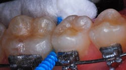
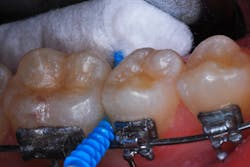
The patient returned for a routine recall appointment on April 6, 2021, wearing fixed orthodontic hardware. A bitewing radiograph was taken (figure 2, top right). The site was cleared with floss, and a new pick soaked with SDF was inserted (figure 3). A small applicator was used to paint sluiceways to enhance the flow of SDF. The pick was slid in and out of the interproximal surfaces a few times (figure 4). The treatment site was immersed in 5% fluoride varnish (figure 5).
After the pick was removed, more varnish was applied. The patient occluded on 2" x 2" cotton gauze for a couple minutes. For the mandibular molar, the pick was inserted above the orthodontic wire in the same way as the maxillary molar was treated (figure 6). After the sluiceways were coated, fluoride varnish was painted over the isolated teeth and the SDF site (figure 7).
The patient returned for a recall appointment on November 24, 2021, 19 months after the initial SDF application (figure 2, bottom).
Other examples include a lingual and double-pick insertion on a teenage patient (figure 8), and a multipick insertion over and under wire placement on another patient (figure 9).
Discussion
There are several brands of dental soft picks available in various sizes. TePe EasyPicks (XS/S-orange and M/L-blue) were used in this case, but other companies market soft picks that can be helpful for this technique (e.g., Sunstar GUM, Practicon Smilegoods PikPak, and others).
The application of fluoride varnish is critical. Besides remineralizing affected enamel, the viscous fluoride coating prevents salivary dilution of the SDF for a few minutes, maximizing its exposure and take-up by the decalcification/caries lesion.
We caution patients that although SDF application attenuates the caries process, caries lesions on contacting surfaces will progress unless daily flossing is part of the oral hygiene routine. We show our patients and parents (caregivers) large, dramatic photographs and radiographs to get that point across.
It’s difficult to standardize contrast on digital bitewing radiographs, so comparative radiolucent changes are equally difficult to interpret perfectly. But in this case, the combination of SDF, daily flossing, initial fluoride varnish, and daily toothbrushing and flossing with a fluoride-containing toothpaste actually “healed” these treated enamel tooth surfaces. The results shown here are typical of what we have observed in patients over the past seven years who are treated with SDF using fluoride dental picks on posterior primary and permanent teeth.
More about SDF ...
Disclosure: The authors did not receive remuneration for this article. Neither author has a financial interest in products or companies mentioned here, with the exception that Practicon Inc. produces and sells Dr. Croll’s education book, The Grossest, Truly Terrifying, but Utterly Awesome Mouth Book.
Editor's note: This article appeared in the May 2023 print edition of Dental Economics magazine. Dentists in North America are eligible for a complimentary print subscription. Sign up here.
References
- Croll TP, Berg JH. Delivery of fluoride solutions to proximal tooth surfaces. Part II: caries interception with silver diamine fluoride. Inside Dentistry. 2017;13(9):56-58.
- Croll TP, Berg JH, Donly KJ. Silver in medicine and dentistry. Inside Dentistry. 2020;16(10):35-40.
- Berg JH, Croll TP. Surface treatments of non-cavitated proximal lesions. Oral Health. 2021;111(12):425-426,428.
- Croll TP, Berg JH. Radiographic verification of silver diammine fluoride action on proximal dental carious lesions in a teenager. Dental Economics. May 6, 2021. https://www.dentaleconomics.com/science-tech/article/14200208/radiographic-verification-of-silver-diammine-fluoride-action-on-proximal-dental-carious-lesions-in-a-teenager


