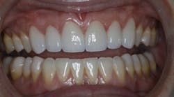
Conservative direct and indirect restorative dentistry to restore vertical dimension and anterior esthetics
Instagram: @joshuaaustindds
Podcast: Working Interferences
The gist:
Posterior composite onlays are used to conservatively open the vertical dimension, allowing for proper restoration of the anterior teeth with full-coverage ceramics.
Kate presented to my practice in the summer of 2020 for a comprehensive examination and our “new-patient experience.” The chief complaint she listed on her new-patient paperwork was, “I am overdue for my cleaning and want to take advantage of my insurance.”
During my patient interview, I noticed that Kate had thin facial enamel on her anterior teeth. It wasn’t until I did my intraoral examination that I noticed the full extent of her issues (figure 1). She had severe enamel erosion on the linguals of her maxillary anterior teeth. After some questioning, she admitted to being a former bulimic who has been in counseling and therapy for several years. She reported that she has been successfully treated and considered this condition to be in her past. As part of our new-patient experience, we take an intraoral scan with our iTero Element 5D scanner. We review those scans with every new patient. Kate’s scan showed the following (figures 2–5). Viewing this scan was the first time that Kate ever had a look at the lingual of her anterior teeth. She was alarmed at the loss of tooth structure and immediately became interested in treatment for this condition. Once a patient expresses an interest like this, we
Kate’s mandibular teeth were largely unrestored. After seeing her scan and photos, she wanted a treatment plan. Now comes the fun and creative part of our job—treatment planning cases like this.
When a patient has wear and/or erosion on anterior teeth (figure 7), we are left with three global options to restore them.1 First, we can use orthodontics to intrude the upper teeth and make room for our restorative material. Second, we could have functional crown lengthening surgery to create more tooth to use for retention and resistance forms. Third, we can open the vertical dimension of occlusion to make space for our restorative materials.
Once Kate saw this, she understood the rationale for treatment and was ready to discuss finances. Generally, treatment plans that include opening of the vertical dimension of occlusion are costly and use a great deal of ceramics. In Kate’s case, I kept thinking about her unrestored lower arch and how I would hate to have to prep away all of that virgin enamel for ceramics. Since she had so much enamel, I thought about possibly opening her vertical dimension of occlusion with resin composite. This would keep both the cost and preparation conservative, as well as allow us the space we needed in the anterior for ceramic restoration with minimal preparation.
The patient agreed to the treatment plan, and we were ready for a more definitive diagnostic wax-up. I sent scans and photographs to Oral Arts Dental Lab in Huntsville, Alabama. I requested an additive diagnostic wax-up on the mandibular posterior teeth to allow an anatomic esthetic wax-up of teeth 6–11 (figure 10). The lab’s Select Department mounted the case and returned the wax-up (figure 11).
On the printed duplicate of the lower wax-up, I made a Copyplast tray using a MiniStar from Great Lakes Orthodontics (figure 13). Using this tray, we made direct posterior composite overlays for the patient’s mandibular teeth (figure 14). This was done using heated composite (Exquisite Restoration Nanofill from Vista Apex), PTFE tape for isolation, and a total etch with universal bonding agent (Scotchbond Universal from 3M).
Once the direct posterior composite overlays were completed, Kate’s occlusion was as it appears in figure 17. We now have the space we need to prepare the maxillary anterior teeth without further reducing the lingual (figure 18).
The preparations were scanned with the iTero Element 5D scanner (figure 19). Provisionals were fabricated with Tuff Temp Plus by Pulpdent (figure 20). The case was sent to the Oral Arts Dental Lab Select Department to fabricate e.max crowns (Ivoclar) on teeth 6–11 (figure 21). Three weeks later, the crowns wereKate was ecstatic with this result, but we are not completely finished with her treatment plan. We have planned to do minimal preparation veneers on teeth 5 and 12, followed by a referral to a periodontist for CT grafting on the lower arch.
This case is an excellent example of using digital direct and indirect dentistry to achieve a conservative treatment plan that answers the patient’s concerns both expeditiously and affordably. Kate would have not been interested in this treatment if we had not performed a scan using our iTero Element 5D at her new-patient experience. Utilizing digital technology at this visit and in our hygiene rooms helps us better communicate the patient’s needs and oral condition. The better patients understand this, the more likely they are to say yes to treatment. Utilizing an intraoral scanner in this way will increase your case acceptance and allow you to do more dentistry!
Reference
- Robbins JW, Rouse JS. Global Diagnosis: A New Vision of Dental Diagnosis and Treatment Planning. Quintessence; 2016.
JOSHUA AUSTIN, DDS, MAGD, is a graduate of the University of Texas Health Science Center at San Antonio School of Dentistry. He is a Pearls for Your Practice columnist for Dental Economics, in which he offers a fresh approach to the critique of dental products and technology. Dr. Austin uses every product that he writes about on patients in his practice, and then gives an honest evaluation for readers. A national lecturer, Dr. Austin maintains a full-time restorative dentistry private practice in San Antonio, Texas. Contact him at [email protected].


