Dentinal hypersensitivity: Contemporary solutions for an age-old issue
Dentinal hypersensitivity (DH) remains a prevalent yet often underdiagnosed condition that affects a significant portion of the global population, impacting individuals across all age groups. Presenting as a short, sharp, sudden burst of pain, DH can lead to considerable physical discomfort and psychological distress, substantially impairing patients’ quality of life.
For DH to develop, two essential processes must take place: (1) the smear layer must be removed through mechanical, physical, or chemical forces, thereby exposing the dentin (lesion localization or LL), and (2) there must be access to the opening of the dentinal tubules, allowing them to connect to the pulp (lesion initiation or LI).1,2 These processes are driven by both distinct and interrelating etiological factors. The etiological factors that can be attributed to LL and LI include erosion, abfractions, recession, whitening, periodontal disease, abrasions, fractured teeth, diet, personal behaviors, and iatrogenic injury.3
Hydrodynamic theory
Despite numerous studies, the precise pain mechanisms associated with DH are still not fully understood. In the 19th century, Gysi hypothesized that fluid flow within the dentinal tubules could activate pulpal nerves upon stimulation, although empirical evidence to support this hypothesis was minimal at the time.4 Decades after Gysi’s initial research, Brännström and colleagues conducted extensive studies over a 20-year period, ultimately supporting the hydrodynamic theory.4 Among the various hypotheses offered to explain the occurrence of DH, this theory remains the most widely accepted explanation.4,5
The hydrodynamic theory suggests that thermal, tactile, osmotic, chemical, or evaporative stimuli induce fluid movement within the dentinal tubules, initiating a pain response due to the activation of mechanoreceptors sensitive to fluid pressure changes.2 Cold stimuli are generally the most intense and pose the greatest challenge for those affected by DH.2
Anderson and colleagues recently found that hypersensitive teeth have approximately eight times more tubules per unit area and larger tubule diameters compared to nonsensitive teeth.6 This structural difference highlights the complexity of DH, which can cause a spectrum of pain intensity from mild discomfort to severe agony, based on various etiological factors and individual patient experiences.
Prevalence
Epidemiological data indicate that DH affects about 57% of the general population, with a notably higher prevalence (60%–98%) among periodontal patients.7 The condition is most common between the ages of 20 and 40, with a slightly higher incidence in women.7 These statistics underscore the widespread impact of DH on both emotional well-being and overall quality of life.
Management of sensitivity
Managing DH effectively involves both identifying the causative factors and employing appropriate therapeutic interventions. Clinically, the approach to DH has primarily focused on treatment rather than addressing the underlying etiological and predisposing factors.2 This emphasis on treatment is understandable given the overwhelming number of products available to manage DH.2
Oral health education
Implementing oral health education is a viable strategy to mitigate DH. This approach includes nutritional counseling for dietary modifications, patient education on risk factors, and modifying brushing techniques. Additionally, it includes educating patients on the proper use of over-the-counter products used to treat DH. These interventions collectively reduce symptoms by decreasing the porosity of dentin. When at-home desensitizing agents fail to provide adequate relief, patients can benefit from in-office treatments. However, as no single treatment offers universal relief, it becomes essential to explore additional therapeutic options.
Therapeutic options
Over-the-counter products and in-office treatments for DH function either through nerve depolarization, which halts neural signaling to eliminate pain perception, or by occluding dentinal tubules through the creation of a smear layer and plugging of open tubules.
Potassium nitrate is the primary ingredient responsible for the cessation of neural signaling in over-the-counter DH treatments. Clinically proven, potassium nitrate creates a protective layer over time by allowing potassium ions to traverse exposed dentinal tubules and reach internal nerves.5 The accumulation of potassium ions desensitizes these nerves, rendering them unresponsive to stimuli such as cold, heat, tactile, and osmotic changes.5 Popular over-the-counter brands containing potassium nitrate include Sensodyne Fresh Mint and Arm & Hammer Sensitive Teeth and Gums Toothpaste. PreviDent 5000 Sensitive can also be prescribed.
Stannous fluoride (SnF2), another active ingredient used widely in over-the-counter treatments, is highly effective in reducing tooth sensitivity by occluding dentinal tubules. Sn ions strongly bond to mineral surfaces on the tooth, forming a protective layer approximately 20 μm deep, which resists dietary acid challenges and blocks stimuli from reaching nerves.5 One study demonstrated open dentinal tubules were effectively sealed after eight weeks of using 0.454% SnF2 toothpaste, significantly reducing sensitivity.5
Although SnF2 can result in a metallic taste, tooth staining, and diminished taste sensation with prolonged use, these issues are mitigated by the addition of zinc compounds such as zinc phosphate and zinc citrate.5 Popular brands containing SnF2 include Crest Pro-Health Advanced Gum Protection Toothpaste, Parodontax Active Gum Repair Toothpaste, and Sensodyne Rapid Relief.
Bioactive glasses, such as Nova-Min found in products like Nupro Extra Care prophy paste by Dentsply Sirona, occlude dentinal tubules by penetrating and forming a physical barrier. They attract calcium and phosphate, promoting hydroxyapatite crystal formation for long-lasting blockage and DH relief.5
Arginine, an amino acid found in treatments like Colgate Sensitive Pro-Relief, operates through both physical and biological processes. Arginine bicarbonate combines with calcium carbonate to form a paste that plugs exposed dentinal tubules. Biologically, arginine fosters beneficial bacteria that metabolize it into alkali, neutralizing acids and helping to maintain a balanced pH.5 This also contributes to the formation of less acidic dental biofilm, enhancing resistance to acid challenges.5
Casein phosphopeptide-amorphous calcium phosphate (CPP-ACP), a milk protein, stabilizes and delivers bioavailable calcium and phosphate, enhancing calcium-binding sites on the tooth surface.5 This action encourages the formation of a crystalline layer over dentinal tubules, providing added protection.5 Despite its irregularities, this layer maintains minerals in a supersaturated state, improving enamel and dentin remineralization.5 CPP-ACP is found in the MI Paste family of pastes and creams.
Nanohydroxyapatite (n-HA) is structurally similar to the hydroxyapatite that comprises 97% of enamel and 70% of dentin. This calcium phosphate compound mimics the natural mineral composition of teeth, replenishing lost calcium and phosphate in exposed dentin areas. By strengthening and remineralizing the tooth, n-HA effectively reduces sensitivity caused by exposed dentinal tubules. Voco manufactures Remin Pro, a tooth cream that combines n-HA with xylitol and sodium fluoride, giving it bacteriostatic and cariostatic properties. This triple-action paste not only remineralizes and strengthens the teeth but also helps with DH relief.
Sodium fluoride is also found in various over-the-counter products and in-office treatments like fluoride varnish. Fluoride varnishes are highly effective and cost-efficient for in-office treatments aimed at mitigating DH. Sodium fluoride occludes dentinal tubules and facilitates mineral deposition, strengthening and hardening the dentin.5 Fluoride ions block exposed dentinal tubules upon contact with the tooth’s surface, reducing sensitivity.5
Given the extensive range of fluoride varnishes available, it is crucial for clinicians to carefully assess both the active and inactive ingredients of each product. Understanding the role of active ingredients, such as sodium fluoride and calcium phosphate, in dentin remineralization is essential for selecting the most appropriate treatment. Additionally, examining inactive ingredients is important to avoid potential patient sensitivities or allergies, ensuring a safe and well-tolerated application.
Voco’s Profluorid varnish effectively provides relief from DH due to its fast fluoride release (two-to-four hours) and enhanced flow characteristics. It is also free from allergens such as tree nuts, peanuts, red dye, corn, shellfish, egg, soy, milk protein, gluten, triclosan, petroleum, saccharin, and aspartame, making it a suitable choice for patients with multiple food allergies or sensitivities.
For individuals with a rosin allergy, which is present in many fluoride varnishes, liquid varnishes offer a vital alternative. Products like Elevate’s FluoriMax and Voco’s Profluorid L effectively decrease sensitivity by occluding tubules. Profluorid L contains 5% sodium fluoride with calcium deposits, making it suitable for patients with allergies to colophony-based materials. The calcium deposits are less water soluble than sodium fluoride, ensuring a longer-lasting release of fluoride and prolonged relief from DH.
Silver diamine fluoride (SDF) should be considered when looking for effective treatment strategies for DH. Some studies have reported DH relief for up to two years.8 SDF blocks acid diffusion and microbial invasion through dentinal tubules.9 The oligodynamic action of silver inhibits microorganisms, and silver particles cover the peritubular zone, penetrating deeply without adverse effects.9 SDF decreases the surface area of dentin, inhibits demineralization, and prevents collagen degradation, providing effective and lasting relief from DH.9
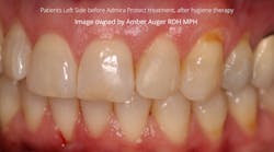
Organically modified ceramics (ORMOCERs) are hybrid polymer materials ideal for dentistry due to their robust inorganic-organic copolymers and inorganic components and have been shown to be an effective treatment option for DH. Voco’s Admira Protect uses nanofilled resin for excellent wear resistance, deep penetration, and sensitivity elimination for up to two years. ORMOCERs are known for their strong adhesion, mechanical stability, and thermal stability.
Lasers are an effective treatment for DH, providing immediate and long-term relief. Lasers work by occluding or narrowing the dentinal tubules, reducing fluid movement. Some lasers also induce hydroxyapatite crystal formation, further aiding in tubule occlusion.10 Both red and infrared wavelength lasers are effective, though effectiveness varies by type.10
Ozone therapy, a newer treatment for DH, requires more study but shows promise. It oxidizes organic debris and bacteria in dentinal tubules, destroying microbes and forming a smear layer that occludes tubules.11 Additionally, ozone increases mineral ion concentration, promoting remineralization and hardening of the exposed dentin surface, thereby reducing sensitivity.11
Final thoughts
There are a plethora of treatment options available for DH, with new methods emerging regularly. This article has highlighted some popular treatments, illustrating the diversity of approaches currently in use. Successful management of DH often requires a comprehensive strategy that addresses underlying causes to prevent recurrence or failure. Unlike caries and periodontal disease, DH management is not always data-driven but relies on logical approaches based on an understanding of its etiology.2
A proposed management strategy involves:
- Accurate diagnosis based on history and clinical examination
- Considering differential diagnoses to rule out other conditions
- Treating secondary conditions contributing to DH symptoms
- Identifying and addressing factors like erosion and abrasion
- Providing dietary advice and oral hygiene instruction
- Tailoring treatments to individual needs based on pain severity and the number of affected teeth2
By integrating these strategies, we can more effectively manage DH and improve patient outcomes.
Editor's note: This article appeared in the September 2024 print edition of Dental Economics magazine. Dentists in North America are eligible for a complimentary print subscription. Sign up here.
References
- Ciucchi B, Bouillaguet S, Holz J, Pashley DH. Dentinal fluid dynamics in human teeth, in vivo. J Endod. 1995;21(4):191-194. doi:10.1016/S0099-2399(06)80564-9
- Addy M. Dentine hypersensitivity: new perspectives on an old problem. Int Dent J. 2002;52(S5P2):367-375. doi:10.1002/j.1875-595X.2002.tb00936.x
- Trushkowsky RD. Etiology and treatment of DH. Decisions in Dentistry. December 8, 2016. https://decisionsindentistry.com/article/etiology-treatment-dentinal-hypersensativity/
- Langenbach F, Naujoks C, Smeets R, et al. Scaffold-free microtissues: differences from monolayer cultures and their potential in bone tissue engineering. Clin Oral Investig. 2013;17(1):9-17. doi:10.1007/s00784-012-0763-8
- Dionysopoulos D, Gerasimidou O, Beltes C. Dentin hypersensitivity: etiology, diagnosis and contemporary therapeutic approaches—a review in literature. Appl Sci. 2023;13(21):11632. doi:10.3390/app132111632
- Anderson CJ, Kugel G, Zou Y, Ferrari M, Gerlach R. A randomized, controlled, two-month pilot trial of stannous fluoride dentifrice versus sodium fluoride dentifrice after oxalate treatment for DH. Clin Oral Investig. 2020;24(11):4043-4049. doi:10.1007/s00784-020-03275-8
- Void-Holmes D. Managing DH with silver diamine fluoride before and after periodontal debridement. SDI. 2020.
- Knight GM, McIntyre JM, Craig GG. Ion uptake into demineralized dentine from glass ionomer cement following pretreatment with silver fluoride and potassium iodide. Aust Dent J. 2006;51(3):237-241. doi:10.1111/j.1834-7819.2006.tb00435.x
- Shah S, Bhaskar V, Venkataraghavan K, Choudhary P, Mahadevan G, Trivedi K. Silver diamine fluoride: a review and current applications. J Adv Oral Res. 2014;5(1):25-35. doi:10.1177/2229411220140106
- Saluja M, Grover HS, Choudhary P. Comparative morphologic evaluation and occluding effectiveness of Nd: YAG, CO2 and diode lasers on exposed human dentinal tubules: an invitro SEM study. J Clin Diagn Res. 2016;10(7):ZC66-ZC70. doi:10.7860/JCDR/2016/18262.8188
- Karlsson L, Kjaeldgaard M. Ozone treatment on dentin hypersensitivity surfaces – a pilot study. Open Dentistry J. 2017;11:65-70. doi:10.2174/1874210601711010065
About the Author

Joy D. Void-Holmes, DHSc, BSDH, RDH
Joy D. Void-Holmes, DHSc, BSDH, RDH, is a leader in oral health care with nearly 30 years of experience. As the founder of Dr. Joy, RDH, and cofounder of JELL-ED, she advances dental education through innovative courses and instrumentation workshops. Dr. Joy holds degrees from Howard University, the University of Maryland, and Nova Southeastern University.

