Pretty as a picture
For more on this topic, go to www.dentaleconomics.com and search using the following key words: photography, imaging, intraoral cameras, technology, Dr. Terry L. Myers.
Photography is a technology that I constantly count on to enhance my practice. American photographer Edward Weston said, “My own eyes are no more than scouts on a preliminary search, for the camera's eye may entirely change my idea.” His philosophy also rings true for photography in my dental office. With so many photography options at my fingertips, peering into the patient's mouth with just a mirror seems to me like only a preliminary search. My camera often changes not just my ideas, but my patient's as well.
Especially for cosmetic cases, the camera is a wonderful way to increase treatment plan acceptance. When a patient is lying down in the chair, I can only see his/her dentition from one angle. In this position, I cannot achieve the full representation that I could if he or she were standing or if I had the opportunity to take a full–arch picture.
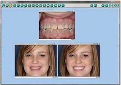
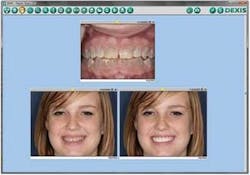
For cosmetic cases, I turn to a Nikon D200 digital SLR camera. With a digital camera, I can get more of the field of view in focus. After taking the photographs, we prepare a PowerPoint–type presentation with the photos on the computer to give the patient an idea of how his or her appearance would change with my proposed treatment plan.
Dental Economics® recently ran a column on Dr. Gregory Lutke and Dallas Dental Solutions. He has become my mentor regarding photography in my practice.
After taking images, I send them to a former dental assistant of mine, Jennifer Moser Johnson at Line Star Dental Arts Studio, where she uses her imaging technology to produce a presentation set to music depicting the patient now, during, and after treatment. This makes it clearly evident where the problems are, what changes need to be made, and what the patient will look like at various stages in the process. The presentation is available so the patient can show it to family and friends. This is a powerful tool for cosmetic cases and practice building.
Besides cosmetic applications, I use my intraoral cameras (DEXIS DEXcam™ 2, DEXcam™ 3; Gendex GXC–300™) many times every day. The easy–to–capture intraoral view gives me a clear perspective of deteriorating amalgams, teeth that may need crowns, or tooth fractures. I can enlarge the image many times over to detect the smallest crack that needs to be treated or watched. An intraoral camera improves my patient's visualization and education. We can have a close–up look at built–up calculus, or I can drop my probe down into the sulcus to demonstrate to the patient the severity of the perio situation. I can print out the image of the cracked tooth or broken cusp for the patient to take home and put on the fridge as a constant reminder to have the tooth fixed soon.
Cameras are such a great decision–making technology with so many attributes: a true view of the present condition for patient education and treatment planning; a great estimation of the outcome after treatment; and intra– and extraoral images from start to finish for documentation of the whole process.
Print them, burn them to a CD, or e–mail the images for patients to have at home — and others will see and want to explore their possibilities as well. Try making photography a part of your technology toolbox. It's a snap, and one more step toward building a picture–perfect practice.
Dr. Terry L. Myers is a fellow in the Academy of General Dentistry and a member of the Academy of Cosmetic Dentistry and the Dental Sleep Disorder Society. He has a private practice in Belton, Mo. You may contact Dr. Myers by e–mail at office@keystone–dentistry.com.

