by Dr. James Kulid
For more on this topic, go to www.dentaleconomics.com and search using the following key words: quality endodontics, cost effective, dental operating microscope, Twisted File, radiology, diagnosis, anesthesia, Dr. James Kulild, Focus On.
The dental world has become an even smaller place, especially during the past 20 years, both in the marketplace and the dental office. The leaps and bounds in technology have been exponential and have spread globally. Information spreads instantly through the Worldwide Web, through Web pages and electronic messaging. Gone forever are the days when dentists could stay aloof from all the new scientific information. Moreover, electronic full-text and full-color PDF files of scientific journal articles are now instantly available to nearly everyone through ubiquitous search portals.
Many of these advances, including the equipment and materials used in these new methodologies, can be used to achieve predictably successful endodontic outcomes. Coupled with these advances are improved quality-of-life issues for both the dentist and patient, allowing for more comfortable treatments with less postoperative discomfort, which increases patient satisfaction. There are also increased efficiencies in practice management, which provides personal satisfaction for the dentist, as well as a high return on investment. These advances extend across the spectrum of endodontic diagnosis, nonsurgical and surgical treatment, and re-treatment.
Diagnosis
Accurate diagnostic methodologies and instrumentation to access pulp vitality have always been crude, since the pulp is completely enclosed in a noncompliant environment and surrounded by dentin. In spite of many attempts to develop instrumentation, which would allow a practitioner to accurately diagnose the disease or health status of the pulp based on the health of the vascular supply, the operator must still rely on diagnostic tests to determine neural response.
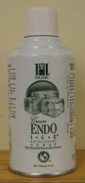
Ice has never been a good tool since it melts rapidly in the mouth, causing potential false positives as the cold water from the melting ice comes into contact with adjacent teeth. Carbon dioxide snow is an excellent way to address this problem, but it is expensive and cumbersome. Endo-Ice (Hygenic, Cuyahoga Falls, Ohio) is a much better tool (Fig. 1) since it is very cold (-26° C), very cost effective, and goes almost instantly from liquid to gas phase, minimizing the chances of false positive responses. Soaking a large cotton pellet with Endo-Ice liquid and placing it on the facial surface of a tooth provides an excellent assessment of pulp vitality. Perhaps in the future, laser doppler flowmetry may provide a predictable way to better determine pulpal health, since it would measure pulpal blood flow instead of neural response.
Radiology
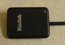
Fig. 2 — Kodak corded CCD radiographic digital sensor
In the past, digital radiography was thought to be a prohibitively expensive toy found only in large multidentist practices (Fig. 2). Silver halide radiographs were still the standard in most dental offices. However, with prices going lower and lower, digital radiographs are found in many more practices, including solo practices. They provide increased radiographic quality as well as cost-effective efficiencies. From a diagnostic perspective, they allow the practitioner to fill up the screen of a 22-inch flat panel monitor with one periapical image. This capability is not only a great diagnostic tool for the dentist, but also a great education tool for the patient.
Software-driven image enhancement tools vary among digital sensor vendors and allow the dentist to change the visual quality of the image, which allows greater opportunities to properly diagnose health or disease. Corded and noncorded digital sensors are available.
With stricter environmental regulations being implemented across the country, dentists can no longer just pour spent developer and fixer down the drain — they must pay someone to dispose of it. Also, the dentist using plain film radiology must continually buy film, developer, and fixer, and buy and maintain an automatic film processor. Additionally, these plain film radiographs must be electronically scanned before being used in any office management software program, or to file electronic claims with third parties.
Anesthesia
Lidocaine has long been the gold standard of local anesthesia in endodontics. It provides profound anesthesia and allows for comfortable manipulation of extremely sensitive pulp tissue. Even though other amide anesthetics, like bupivocaine, provide longer lasting anesthesia, they do not provide the profound degree of anesthesia afforded by lidocaine. However, a relatively new amide local anesthetic called articaine is poised to take the place of lidocaine based on its rapid onset and ability to provide even more profound anesthesia.
It is a 4% solution versus the 2% found in lidocaine, but the 1:200,000 concentration of epinephrine in articaine is less than the 1:100,000 in lidocaine. Current evidence indicates there is no increased risk of paresthesia with articaine. If a patient has a documented allergy to amide local anesthetics, Nesacaine, an ester, can be purchased in multidose vials and an appropriate concentration of epinephrine can be added.
The dental operating microscope (DOM)
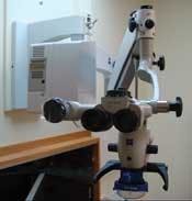
Price breaks on high quality DOMs have put the instrument within reach for most dental practices (Fig. 3). Moreover, its use is not confined to endodontics. A DOM can also be used for restorative dentistry, periodontics, and oral and maxillofacial surgery. The increased magnification also allows for superb documentation of patient treatment for insurance submission, patient education, and educational presentations for fellow health care professionals.
Fig. 3 — Wall-mounted Zeiss OpmiPico five-step dental operating microscope with Zenon light source
Not only does the DOM substantially increase the functional capabilities of the dentist, it tells patients that their dentist is practicing the latest state-of-the-art dentistry. High quality endodontic treatment cannot be accomplished without enhanced magnification technologies. It's not used during all phases of treatment, but its use is critical at some point in nearly all cases to achieve high rates of healing. Although floor-mounted models are excellent, wall- or ceiling-mounted models are easiest to use.
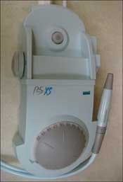
Ultrasonic instrumentation is also critically important to quality access preparation (Fig. 4). It can be used with the DOM and does not occlude the field of view through the eyepieces of the DOM like high- or low-speed handpieces. An extremely wide variety of ultrasonic tips are available, which allow for wide flexibility when removing precise amounts of dentin while locating calcified RCSs, or disassembling posts and cores, etc. Plastic ultrasonic tips are also more cost effective than traditional stainless steel or titanium tips.
Nonsurgical root canal treatment
It wasn't too long ago that endodontists learned their specialty in much the same way that Dr. Louis Grossman learned it early in the 1900s: arduous hand-filing and reaming followed by cold lateral compaction (CLC) of gutta percha or placement of silver cones. But those days are gone.
Fig. 4 — Satelec ultrasonic instrument (Acteon) with BUC 1 tip inserted
Even though hand files are and will continue to be an integral part of any quality endodontic treatment, more flexible and efficient nickel-titanium (NiTi) rotary instruments have now assumed the dominant role in the cleaning and shaping of the root canal system.
As a safety measure, hand files should always be used to enlarge root canal systems to at least a size No. 20 before introducing any NiTi rotary file. This step will minimize the chances of small NiTi rotary files separating. In the past, all NiTi files were ground to create their shape, unlike hand files that were traditionally twisted from either square or triangular blanks of stainless steel wire. However, NiTi files are also now produced through stamping and twisting.
Stamping is used to make the LightSpeed LSX file (Lightspeed, San Antonio, Texas), a completely new design. The new instrument retains the same basic design feature that produces a round preparation at the most apical extent of the instrument and apical root canal preparation.
As a result, the dentist must use another instrument that produces a flaring of the root canal system, which allows for maximum cleaning and shaping, and produces a shape that allows for obturation of the prepared space with an appropriate material and technique.
The new Twisted File (SybronEndo, Orange, Calif.) is produced through a totally new manufacturing process that uses proprietary equipment and techniques. The process of twisting the files allows for the microcracks and surface irregularities to be aligned along the long axis of the file.
This is in contrast to the traditional ground NiTi file, where those surface irregularities are aligned perpendicular to the long axis of the file, which allows the possibility for increased risk of instrument separation during use.
However, electropolishing of traditional ground NiTi files decreases the sizes of these imperfections. There are few evaluations of these new instruments. But initial reports indicate they have promise for efficiency and quality; however, they are more costly than traditional ground NiTi files.
How many times should a practitioner use an individual NiTi file in an RCS, no matter how it is manufactured? Reports indicate this is more a function of how the operator uses the file rather than the file itself.
One of the most important things a dentist can do to decrease the incidence of instrument separation is to view the file under the operating light before putting it into an RCS and after taking it out. It should be examined for any light reflection irregularities that would indicate a defective area on the file. If one is found, the file should be discarded.
“When in doubt, throw it out” are good words for the wise. No matter what the dentist tells the patient or what informed consent the patient signed, they still don't want to hear that the dentist just “broke a file in my tooth!”
A good way to find out which NiTi system would work best in a practitioner's hands is to ask a company representative for a sample of their product, which can then be evaluated by using it on some extracted teeth in the laboratory. If the system seems promising in the lab and the operator likes it, then it's time to use it clinically to see if it has merit in patient treatment.
Obturation
Gutta percha remains the core material of choice, although much has been written about the pros and cons of Resilon, a resin-based material used in conjunction with Epiphany, a resin-based sealer. Resilon/Epiphany is an excellent system for obturating an RCS. However, it's more expensive than traditional GP systems, and the literature does not report that it's any better in long-term healing, biocompatibility, or sealability.
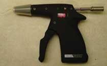
Fig. 5 —Cordless HotShot gutta percha injectable system
The more important consideration is the technique used to obturate the RCS. Although CLC remains the most widely used technique, it has significant shortcomings. Many of these are addressed by thermoplasticized techniques that soften the GP, allowing it to more completely obturate the root canal system with less sealer than is required by CLC. Since sealer is resorbable, it's desirable to minimize its volume.
There are a variety of thermoplasticized techniques available, including injectable warmed GP (Obtura III, Calamus, and HotShot), carrier-based systems (Thermafil, GT, and ProTaper Obturators), warm lateral compaction (Endotec II and DownPak), and continuous wave (CW) (System B, DownPak, and Touch ‘n' Heat). The HotShot (DiscusDental, Culver City, CA) cordless small footprint ergonomic design makes it ideal for a crowded dental operatory (Fig. 5).
The DownPak (Endo Ingenuity, Chicago) enhances the CW technique with its cordless design and adjustable temperature settings (Fig. 6). Also, the operator can use the vibration capability of the instrument to help thermoplasticize the mass of GP.
No matter the technique used to clean, shape, and obturate the RCS, the timely placement of a definitive restoration over the completed RCT is critical for optimal healing.
If a temporary restoration is placed initially, it should be replaced by a definitive restoration or core buildup within three weeks. It takes little time for a superb endodontic obturation to become contaminated if exposed to oral fluids, which would then require a subsequent re-treatment.
Ultimately, our task as skilled dental health care professionals is to render predictable quality dental care to our patients. The operator and patient are both better served when the dentist chooses to do procedures in a skilled way for which he/she has the training, desire, experience, expertise, and armamentarium. It makes sense to select those procedures where the return on investment (time, equipment, money, and training) is high.
The Case Difficulty Assessment Form (www.aae.org) of the American Association of Endodontists is an excellent tool to help identify cases where the dentist can be predictably successful — measured in both positive healing outcomes as well as income.*
Selecting the right cases can make that happen. Referring the more difficult cases can free up time in a busy office to accomplish those procedures where the ROI is more predictable, versus spending a prohibitively expensive amount of time attempting to locate calcified RCSs, establishing access preparations through crowns, re-treating contaminated cases, or attempting difficult endodontic surgical cases.
Endodontic treatment results in predictably successful healing outcomes. Further advances in armamentarium and techniques will allow endodontic therapy to enjoy even higher success rates, which allows patients to retain their natural dentition.
Dr. James Kulild earned his DDS from the UMKC School of Dentistry, an MS in oral biology from George Washington University, and dndodontic training at Walter Reed Army Medical Center. He is a diplomate, American Board of Endodontics, and secretary, American Association of Endodontists. He is professor and director, postgraduate dndodontics, at the UMKC School of Dentistry. Contact him at [email protected].
Editor's Note: Fig. 7 for this article is available on the Dental Economics® Web site at www.dentaleconomics.com. Click on the Download Center and select this article title.

