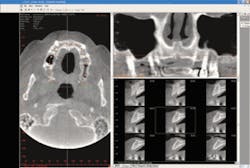
The key to a safer and more accurate diagnosis is just one dimension away.
Cone beam technology is quickly advancing the dental industry’s approach to patient treatment, diagnosis, and outcome predictability. Cone beam technology has never been more valuable, and is changing the way we do dental diagnosis.
One dentist who speaks from experience is Kim Gowey, DDS, a 30-year general practitioner and past AAID president who focuses on implant surgery and prosthetics at Implant Dentistry of Wisconsin. He’s used Imaging Sciences International’s i-CAT® Cone Beam 3-D Imaging System in his office for nearly two years.
We took a bite out of the technology with Dr. Gowey, and drilled into how and why cone beam machines and the i-CAT are revolutionizing his practice and patient care.
DE: How is the i-CAT different from traditional 2-D X-rays?
Dr. Gowey: In my 30 years of dental practice, there’s never been anything like the i-CAT for diagnosis. Cone beam CT is revolutionizing the way treatment-planning is done. With cone beam technology, dentists can perform 3-D diagnoses versus traditional panoramic and cephalometric flat film. Patient images in 2-D are distorted and magnified. These images often make it difficult for the practitioner to locate critical anatomies, pathologies, and other important anatomical concerns because they are not visible in the scan. Cone beam 3-D imaging allows dentists to see a patient’s anatomy in all dimensions by creating a 360-degree analysis. These images allow dental professionals to achieve a complete makeup of the human jaw, face, and associated structures, which leads to greater accuracy, precision, and efficiency in diagnosing and treating patients.
In orthodontics, instead of relying on flat cephalometric and AP views, CBCT 3-D allows asymmetries to be visualized, making diagnosis more accurate. I can view the entire skull on a computer screen, rotate it, and superimpose the soft tissue. For instance, I can see a periapical lesion hiding behind the root that doesn’t show up on a periapical radiograph very well. I can clearly visualize impacted teeth. I can view the inferior alveolar nerve in relation to the roots of the teeth, and can clearly delineate endodontic lesions.
There is also the ability to see calcifications in major blood vessels in the upper neck and visualize sinus anatomy. The temporomandibular joints can be examined in 3-D. Compare this to a tomogram, which gives single slices through a joint so the whole joint can’t be seen. With CBCT, we can examine the bony joint surfaces and shape in detail. Cone beam scanning also benefits patients, exposing them to significantly less radiation than a medical CT scan.
DE: Why did you invest in cone beam technology?
Dr. Gowey: I first saw the i-CAT soon after it was introduced in November 2004 during the American Academy of Implant Dentistry annual meeting in New York. I was amazed by the technology and images. The machine does a 10-second panoramic scan, normal CBCT scans, and also high-resolution CBCT scans. I ran the numbers on how many panoramic scans I perform and how many potential implant scans we would do, and I figured the payback on the i-CAT would be under five years. Now that I’ve had it since June 2005, I think it’ll pay for itself in less than three years.
DE: What was the learning curve?
Dr. Gowey: Learning how to use the i-CAT and understanding the benefits of cone beam technology came very quickly. My staff learned the ins and outs of the machine in just a couple of days. Imaging Sciences installed it in one day and we were trained the next. It is easier to position a patient in the i-CAT than in a panoramic X-ray machine because there is no focal trough. If you do orthodontics, you get a panoramic view, cephalometric view, and CT views from one scan. You can make orthodontic cephalometric measurements right on the computer screen. The images can be viewed in any of your operatories over your network.
DE: What difference has cone beam technology made in your practice?
Dr. Gowey:It has increased case acceptance. With cone beam technology, dental professionals can analyze the complete surface of a patient’s mouth, face, and jaw. The 3-D scan also exposes issues not detected with traditional methods, which gives us better insight into the relationship between patients’ underlying dental structures and soft tissue.
As a result, I’ve been able to find the unexpected well in advance of a procedure, shorten treatment-planning time, and increase diagnostic accuracy and surgical predictability. The i-CAT replaces a cephalometric and panoramic X-ray machine. Since it is digital, the images can be manipulated to change contrast and brightness and apply filters to help you find what you are looking for. There is so much information available to you from one scan compared to conventional radiography. One scan gives you an image of the maxilla, mandible, TMJ, sinuses, nasal cavity, and area back to the cervical spine. You can see root positions in the bone for orthodontic treatment, bony dehiscences and fenestrations, abcesses draining into the maxillary sinus, impacted tooth locations, salivary stone location, and many other pathologies in the maxillofacial area.
Very accurate measurements can be made directly on the images. Bone density can be measured in any area on the scan. TMJ mapping is very easy. A panoramic view is normally mapped and can be done separately for the maxilla and mandible to improve image clarity if needed due to malocclusion or jaw-size discrepancy.
The comprehensive 3-D images are a valuable tool to patients who are considering necessary and elective procedures. It’s especially helpful when a dentist is trying to show patients the value of a procedure. The scans reveal detailed information that’s crucial for implant procedures and third-molar removal, such as locating the inferior alveolar nerve canal to avoid complications. The immediate 3-D digital reconstruction allows us to share a visual diagnosis with patients so they can better understand their treatment options, while exposing them to a significantly lower radiation dose compared to traditional CT scans.
DE: Do you have any specific patient stories to share?
Dr. Gowey:I saw a patient a couple of years ago who was considering placement of two lower posterior implants. Since his ridge was thin and short, we thought he would need a bone graft prior to implant placement. He ended up not having the procedure done. He was in again recently and I said, “Let’s take a cone beam scan so we’ll know exactly what we can do.” I measured his ridge while he watched, and determined he was indeed a candidate for the implants without the need for a bone graft. He said, “It’s really fantastic to see this right here. The bone graft scared me off before, but now it’s no problem, so I’m going to have it done.”
DE: What has been the reaction of patients and staff to the cone beam technology?
Dr. Gowey: They really like it. There’s no dark room, chemicals or developer expense. It’s digital, so they can view the scans right away when I do. Patients are impressed with the new technology and they can see the benefit. They appreciate that we have the latest technology that improves their care. This makes our practice unique in patients’ eyes. They don’t have to make an appointment at the hospital or imaging center for the scan, they can have it done immediately during an appointment, and walk out with the information they came for. They appreciate the convenience and the fact that the fees for CBCT are much less than for a hospital CT.
DE: How often do you use the i-CAT?
Dr. Gowey: My practice scans patients several times each day.
DE: Was it difficult to integrate in your office?
Dr. Gowey: It’s actually the same size as a panoramic machine. The computer sits next to the door adjacent to it. It was a perfect fit and did not require any office changes.
DE: What is the bottom-line impact on your practice, both in terms of finances and patient perception?
Dr. Gowey: I thought our investment paid for itself over a reasonable period of time. The profit comes from increased treatment acceptance. I saw so much value in the information obtained from the machine for my practice and patients, and I continue to benefit from it daily.
It directly influences my practice’s overall quality of care. Patients experience less discomfort postoperatively, and benefit from shorter appointments, faster procedures, more accurate placement, faster recoveries, and cost savings. This is due to the ability to do minimal flap reflection for implant surgery because the bone can be visualized on the computer screen.
A surgical guide from the CBCT data to accurately place implants can be made without extensive flaps. The visual diagnosis helps communicate their conditions so they can better understand their treatment options. The preoperative scans help me perform safer, less invasive procedures by increasing precision and reducing risk for the practice and patients.
DE: What would you tell your colleagues who are interested in cone beam technology?
DE: What would you tell your colleagues who are interested in cone beam technology?
Dr. Gowey:There are a variety of new machines. There are a couple of Japanese ones, but the field of view is small. There’s a European machine that gives cone beam images, but there’s much more scatter in the views, so it’s difficult to make models from these, according to Dr. Jerry Soderstrom, who is an expert on CT and 3-D model-making. The i-CAT is the original, and it’s made in the United States, which I really like. It’s not invasive, it’s simple to use, and it’s convenient for the patient. To me, it’s a no-brainer. It’s too easy to not have this technology available to you.
How i-CAT Works
The Imaging Sciences i‑CAT is the leader in Cone beam 3-D imaging by producing detailed, three-dimensional views of all oral and maxillofacial structures. The i-CAT gives dentists and specialists the most complete information on the anatomy of a patient’s mouth, face, and jaw areas, which leads to the most accurate treatment and predictable outcomes for patients’ surgical procedures.
The entire i-CAT workstation - including machine, computer station, and access space - requires less than 30 square feet of office space. It creates 3-D images at a reduced cost and with considerably less radiation than traditional Fan Beam Computed Tomography systems.
Patients remain seated for just 20 seconds in an “open environment scan,” which increases patient comfort and captures the natural orientation of anatomy. The data is transferred to a computer within minutes, and displayed on an intuitive 3-D mapping tool that lets doctors and technicians easily format and select desired “slices” for immediate viewing.
The i-CAT is used as part of the full scope of dentistry, including oral and maxillofacial surgery, impacted teeth, implants, bone reconstruction and grafting, orthodontics, trauma, pathology, screenings for infection, and TMJ evaluation. Visit www.i-CAT.com for more information.


