Dr. Spurgeon Webber
by Jeffrey B. Dalin, DDS, FACD, FAGD, FICD
Dr. Dalin: Spurgeon, from doing a little research about you, I see that we have much in common. We are both huge fans of dental technology, and we use the newest technologies to deliver the best dental care to our patients. I think discussing this subject will help our readers get a better understanding of how to integrate these technologies into their practices.
Dr. Webber: Thank you, Jeff. It is a pleasure to discuss some of the uses of new technologies in the dental field.
Dr. Dalin: Let’s start with one of the basics ... magnification. One of the biggest challenges during many of our procedures is to visualize what we are working on. An example of this is the detailed margins of a crown. These are only as good as the detail that is visible to the clinician. Dental loupes allow the operator to see more, and ensure an excellent end result. I know we have many types from which to choose.
Dr. Webber: Magnification is one of the most important and underutilized technologies that doctors should have in their arsenals of tools to deliver their best possible dental care to patients. It is a part of our treatment modality that benefits the doctor as much as the patient.
Dr. Dalin: I know practitioners, including hygienists, can benefit from magnification. I would suggest going to a meeting, talking to vendors there, and trying out their products, then choosing what works best for you. But I know that magnification is not the only factor involved in better visualization. Are you a fan of adding portable LED light to enhance vision?
Dr. Webber: I am a huge fan of portable LED lighting, no matter whether these are placed on loupes or instruments, like handpieces, mirrors, or oral isolation devices. Anything that betters visualization of the oral operational structure or site improves treatment results.
Dr. Dalin: I understand that you have a MagnaVu scope. Tell us about this technology.
Dr. Webber: I tested various magnification scopes, and decided on the MagnaVu as the best fit for my practice because of its ease of operation and field of view. I think the MagnaVu system is better adapted for the general practitioner. I was confined by dental microscopes and discovered that I had more flexibility and less eye fatigue with the heads-up display on a high-definition monitor.
Dr. Dalin: How does this scope differ from a dental microscope?
Dr. Webber: The dental microscope is a viable and useful system; however, it is different from the MagnaVu system. Instead of working through a viewfinder, you work while looking at a high-resolution monitor. MagnaVu uses a high-resolution digital color CCD camera chip. When combined with the company’s custom optics, it produces clear, precise images. It has an incredible depth-of-field (up to four inches). This means that everything in the mouth can be in focus simultaneously. Depth perception is no longer an issue. The MagnaVu features a fully adjustable custom lens system that allows the user to choose the desired optical magnification level from 1X to 23X. Imagine being able to view images 400 times larger than normal. The versatile MagnaVu can look down a root canal, be positioned for quad dentistry, display an entire arch for hygiene procedures, or retract to take beautiful full-face images of a patient’s smile.
Dr. Dalin: Another plus of the MagnaVu system that people often don’t think about is helping posture and avoiding back and neck health issues.
Dr. Webber:We are taught in dental school the correct posture when treating patients. Over time, we tend to bend our heads periodically for direct visualization of procedures. This can have dire consequences for neck and back discomfort. Just as the proper use of a mouth mirror with correct posture can prevent neck and back problems, MagnaVu assists by allowing the doctor to move the camera and patient rather than the dentist’s neck and head.
Dr. Dalin: While we are talking about visualization, I still cannot understand why 100 percent of dental offices do not utilize digital radiography. Could you imagine practicing without it?
Dr. Webber: I could not imagine practicing without digital radiography today. Besides its safety and environmental benefits, patients easily can see the radiolucencies and radiopacities that warrant our treatment plans. Information is obtained in seconds as opposed to minutes, making for exponential increases in practice efficiencies. Though the initial cost might appear high for a digital X-ray system, you recoup the expense in less than a year if you see as few as five new patients or periodic exam patients per week. In addition, insurance companies cannot deny the treatment because they might have lost any X-rays. We can send them as many copies as necessary.
Dr. Dalin: What impact has the CEREC CAD/CAM had on your practice?
Dr. Webber: Again, we are talking about exponential increases in efficiency in delivery of care to our patients. Digital radiography gives us exponential increases in speed and amount of information from a diagnostic standpoint. CEREC gives the doctor exponential increases in efficiency of delivery of actual treatment for our patients. It used to take days to weeks to get a crown. Now it just takes minutes to hours to get a crown. When I began using the system in 2000, I received resistance from an insurance company because it could not understand how my patients’ claims had the same crown prep and crown seat dates. After sending the company research information and an invoice as proof of purchase of the technology, there were no further delays of claim submissions.
Another benefit of CAD/CAM is near real-time feedback on esthetics and occlusion. Changes can be made on the computer to minimize intraoral refinements. If, for some reason, material or design fails, you can make the correction on the spot.
Dr. Dalin: How do you utilize hard- and soft-tissue lasers?
Dr. Webber: Since 2001, I have used the hard- and soft-tissue Biolase laser. It gives doctors additional tools in their arsenals to comfortably and efficiently care for patients. Anything that can be done with a bur or scalpel can be achieved with a hard- and soft-tissue laser; however, there are some procedures that can be done with a hard- and soft-tissue laser that cannot be performed with a bur or scalpel.
For example, one can set a laser to remove decayed tooth structure while preserving healthy tooth structure. Patients appreciate this minimally invasive approach to dental treatment. You also can set the laser to make soft-tissue incisions with minimal to no bleeding while sterilizing the site as you laze. These procedures can be done with no to minimal local anesthesia. Patients appreciate a shorter chair time not having to wait for an anesthetic to take effect. Patients also like getting no or less anesthetic. This negates or minimizes the lingering numbing effects after procedures are complete.
Dr. Dalin: I know dentists can be hesitant about adding new equipment and procedures, or not understanding how to use these procedures properly. You must have a plan ... from undergoing training to adding the new procedures. If used properly, these technologies will pay for themselves. Can you offer any suggestions to our readers about making this step?
Dr. Webber:Each doctor who is looking to add new technologies or systems to his or her practice will have different needs. The constants in dental practices are administrative, diagnostic, and treatment system needs. A computer network with good practice-management software should be the foundation of any new technology plan. From there, digital radiography and digital imaging will form the basis for diagnostic systems. Treatment systems, like CAD/CAM and lasers, should be added in phases based on budgets, formulas of need, and productivity for individual practices.
Dr. Dalin: Is there anything else you would like to discuss?
Dr. Webber: Technology alone does not make a good doctor. Technology allows good doctors to practice better dentistry by better informing their patients to treatment recommendations and by performing that treatment more efficiently, predictably, and more comfortably. This, in turn, makes the doctor’s experience healthier and more enjoyable.
The new technologies complement most of the existing, older technologies; however, they do not make previous dental technologies obsolete. Mirrors, explorers, probes, and cotton pliers continue to comprise our basic setup. We continue to use handpieces, scalers, and curettes well into the 21st century. Our dental labs are indispensable. They are incorporating these technologies in case fabrications.
Digital imaging with laser diagnostic tools, such as DIAGNOdent and digitized light caries detection, make it important for the doctor to digitally document findings.
There is resistance from some insurance companies to not pay for restorations unless radiolucencies are seen on a radiograph.
Ethical doctors know that there are many cases in which decay that warrants treatment may not appear on a radiograph. It is important that we begin to record site size classifications of caries in our notes along with digital photographic records of lesions that are visualized yet not apparent on a radiograph. The digital photographic series should be part of the digital radiographic series of images for our patients.
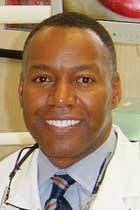
It is important that doctors research and test the technologies that they plan to add to their practices. If possible, do a trial run of the technology in your practice, or visit with other doctors who use the technology. Once the decision is made on a particular technology, commit to it and become well-versed in its application.Dr. Spurgeon Webber III has been in private practice for 21 years in Charlotte, N.C. He is past president of the Charlotte Medical Dental Society, and lectures on dental systems and technologies that increase practice efficiency and productivity. Dr. Webber graduated from Duke University, and received his DDS from Howard University College of Dentistry in 1986. He is also on staff at Carolinas Medical Center in Charlotte. For contact information, send an e-mail to [email protected].
Jeffrey B. Dalin, DDS, FACD, FAGD, FICD, practices general dentistry in St. Louis. He also is the editor of St. Louis Dentistry magazine, and spokesman and critical-issue-response-team chairman for the Greater St. Louis Dental Society. He is one of the co-founders of the Give Kids A Smile program. Contact him by e-mail at [email protected], by phone at (314) 567-5612, or by fax at (314) 567-9047.
