New tools for oral cancer
The eonomics of saving lives
by Ken A. Neuman, DDS
Isn’t it strange to talk about economics and saving lives at the same time? That is exactly what dentists will be doing with two new devices - VELscope and ViziLite - now available in the dental market. Many of us will base the decision on whether or not to acquire these new devices on economics. If economics is the basis of your decision, ask yourself how valuable this technology would be if it saved the life of a friend, family member, or son or daughter.
Will one of the two technologies fits your practice? I am convinced one of the two will! My real challenge in this short article will be to convince you that one of these two devices needs to be in every dental office, and that you can afford to pay for the device of your choice very soon after acquiring it. I would like to say that regardless of cost, you should have it and use it, but I know that the economics in any technology must be a consideration.
VELscope
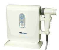
Through the use of a light shining on the intraoral tissues to induce tissue fluorescence, the VELscope (LED Dental Inc., White Rock, BC) assists in the detection of abnormal areas - including precancerous lesions - sometimes before they become apparent under white light examination.
VELscope was designed to be easy to use and provide the general practice with an adjunctive method to help determine when further evaluation is needed. We have been doing visual and tactile oral cancer exams for many years. When I first used this device, it opened my eyes to how much more we could do and how much earlier we could catch incipient lesions, refer patients for further investigation and evaluation, and in doing so, be on the side of the curve that has my patients surviving oral cancer.
Estimates are there will be nearly 31,000 new cases of oral cancer and over 7,400 deaths from it in the U.S. in 2006 alone. According to the American Cancer Society, the most common cause of this high death rate is that lesions are discovered too late.
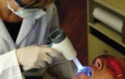
VELscope works by illuminating tissue with a beam of blue light of a specific bandwidth, which excites the tissue from the surface to the basement membrane (where pre-malignant changes typically start) and into the stroma beneath. As a result, the excited tissue produces its own light (i.e., it fluoresces). Through proprietary optical filtering in the VELscope handpiece, the clinician can see the resulting fluorescence and the changes in fluorescence that occur when abnormal tissue is present. Healthy tissue typically appears as a bright green glow, while abnormal tissue can cause a loss of fluorescence and thus may appear dark. In a clinical study conducted by the British Columbia Cancer Agency, which involved 44 high-risk patients, VELscope achieved a sensitivity of 98 percent and a specificity of 100 percent when discriminating between normal tissue and severe dysplasia, carcinoma in situ (CIS), or invasive carcinoma, using histology as the gold standard.
The method used is very simple. After performing your standard visual and tactile mucosal exam, you simply re-evaluate the tissue by looking through the VELscope Handpiece. The fluorescence visualization will assist you in distinguishing between healthy and suspicious tissue. The VELscope exam takes less than two minutes, and there are no extra steps required, other than turning the device on and viewing the intraoral tissues through its handpiece.
ViziLite
The second device we will discuss is the ViziLite from Zila Pharmaceuticals, Inc., in Phoenix, Ariz.
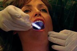
Figure 3 shows a photo of the ViziLite Plus being used. The patient must first rinse with an acetic-acid mouth-rinse for 30 seconds to remove the mucoproteins. Then, the clinician activates a disposable chemiluminescent light stick. Once activated, the light stick is then used to illuminate the oral cavity. If an area of concern is detected, TBlue 630 - a toluidine blue metachromatic dye - is used to stain the tissue in the oral cavity and the light must be used again. Subtle abnormal changes in tissue may appear blue in the presence of the ViziLite.
Doctors, you have the potential to save lives. Dr. Miriam Rosin, director of the BC Cancer Agency’s Oral Cancer Prevention Program, has stated that “When oral cancer is detected early, patients have an 80 percent chance of survival.”
Dr. Ken Neuman is an accredited member of the American Academy of Cosmetic Dentistry. He works with Dr. Gordon Christensen as a CRA evaluator. He has earned Fellowships in the Academy of General Dentistry, the Academy of Dentistry International, International College of Dentists, and American College of Dentists. Contact Dr. Neuman by e-mail at [email protected].


