State-of-the-art 2-D and 3-D implant planning
With the increasing popularity of dental implants, doctors face a myriad of dental conditions that can affect the implant’s viability. To gain a closer look, dentists have moved toward the CAT scan (3-D) imaging for treatment planning. There is another valuable option that takes advantage of 2-D imaging common in a growing number of dental practices - the digital intraoral and panoramic X-ray.
I’ve asked my colleague, Dr. Robert Fazio, a private-practice periodontist and associate clinical professor of surgery at Yale University, to offer some insights into 3-D and calibrated 2-D imaging.
Brad:Robert, how do you approach 3-D and 2-D imaging in your implant planning?
Robert: As part of the planning process, I evaluate a patient based on clinical parameters, inspection, and palpation of the edentulous ridge, tissue sounding, and other intraoral findings clinically, combined with the use of conventional radiographs. In cases with less available bone, it is imperative to have more information to avoid violating vital structures like the maxillary sinus or inferior alveolar nerve.
CAT scan technology provides a very specific three-dimensional view of the bone without the distortion that occurs on panoramic film and somewhat on periapicals. 3-D provides the ideal image for implant treatment. There are times when availability is limited. For these situations, calibrated 2-D imaging can offer another treatment option. Initially, it can expedite the diagnosis and treatment planning for any patient, and often provides enough information to complete the diagnosis.
Brad: What is entailed with this calibration process?
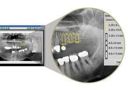
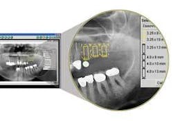
Robert: We can clinically, in a matter of seconds, place a 5 mm steel ball held in place with simple wax and shoot a conventional digital pan or periapical with our digital radiography software. Using the same program’s implant tools, we click on that ball on the image to allow the software to adjust for distortion. Then we drag-and-drop the desired implant model per specific manufacturer onto the site. It appears accurately sized with safety margins. This is an extraordinary way to increase the speed and thoroughness of the diagnosis.
Brad: How does this improve communication with colleagues?
Robert: It provides for maximum communication between the restorative dentist and whoever is placing the implant. Overcrowding can result from the quest by the surgeon to get a larger, wider implant or multiple implants into the space to achieve more support for the final restoration. Using the software’s implant functions, the restorative dentist may literally demonstrate spacing that is inappropriate for larger implants. The dental team is now planning the ideal restoration even before the implants are placed.
Brad: You mentioned safety. How does this implant imaging software facilitate this?
Robert: The software’s library is implant-specific. Regardless of the implant system the surgeon uses, he or she can specify the exact model of the implant and “drop it” into the exact location on the calibrated radiograph. Around those images there is a 1.5 mm “safety box” that allows you to space the implant safely and predictably from vital anatomical features and other implants.
Brad: How do you educate patients about the procedure?
Robert: Nothing places the patient more at ease than seeing a radiograph schematic of the implant with the safety margin drawn in on the image. Patients can take this home for later reference.
Brad: Is this process cost-effective?
Robert: Investing in this module is substantially less than the cost of placing one implant - inconsequential to anyone doing even a handful of implants on a yearly basis.
Brad: Thanks, Robert, for sharing this information.
Robert: Glad to be of service.Dr. Brad Durham has practiced dentistry for 25 years with an emphasis on the treatment of head, neck, and facial pain, dental cosmetics, and complex dental reconstruction. His practice combines art, science, and technology with personalized care. He is a clinical and featured instructor at the Las Vegas Institute, and was the first in the world to earn the LVI Mastership award for esthetic reconstruction. Dr. Durham teaches a series of courses titled “The Niche Practice” at LVI and his home in Savannah, Ga. Contact him at [email protected] and www.nichepractice.com.

