CAD/CAM and 3-D Vision Beyond expectations, both clinically and in business
By Neal Patel, DDS
It is well known that I hang my hat on CBCT. With the introduction of CBCT to dentistry, I have observed a giant leap in my diagnostic ability. It is the cornerstone of my practice. I also feel fortunate to practice dentistry in a period that has witnessed the growth of CAD/CAM. The rise of digital dentistry is led by CAD/CAM, allowing clinicians to image, design, and mill restorations chairside in one visit. I have come to appreciate and rely heavily on CEREC as a restorative tool. Together, these technologies allow me to provide an incredible level of comprehensive care to my patients.
I once hesitated to recommend these technologies to other clinicians unless I really knew who they were and what kind of dentistry they were practicing. In particular, I was fearful of having to look at the "sticker-shocked" faces of my colleagues. Today, I recommend these things to them without hesitation, asking only that they invest in education to maximize the use of the technology.
Today, I enjoy the "sticker-shocked" faces. I show them my practice numbers, and I enjoy watching their dumbfounded expressions as eyes pop and jaws drop! You pay to play when it comes to these technologies. Both CAD/CAM and CBCT have fantastic track records in supporting clinicians both clinically and in business.
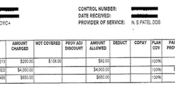
In particular, CBCT has opened opportunities for my practice to bill procedures to medical insurance! As a teaser to keep you interested, here is the medical insurance reimbursement for the exam, CBCT, and OSA appliance alone for the patient at the end of my article. (FIg. 1)
Yes, you read this correctly: a total of $4,742 paid for by medical insurance. This does not include the extraction, bone graft, subsequent CBCT, and surgical guide … or endosseous implants also covered by medical insurance! I am sure I have your attention now, so let's move forward.
Combining CAD/CAM and CBCT in a single practice with complete integration creates a magical experience for the clinician and the patient alike. CBCT provides an opportunity to provide an objective assessment based solely upon results of the 3-D image. With the rise of technology within society, today's patients demand the most advanced dental care. 3-D diagnostic images help us treat patients with proper diagnosis. They also lead to higher treatment plan acceptance and increased precision in dental therapy.
The integration of CBCT diagnostics with CAD/CAM and other objective data from biometric instrumentation, like the SICAT JMT (Jaw Motion Tracker), gives us an opportunity to formulate a definitive treatment plan with a common goal: optimal oral health.
The biometric instrumentation provides clinicians the opportunity to evaluate dysfunction in the temporomandibular joint, the craniofacial musculature, and the overall stomatognathic system. Use of Sirona's Galileos (CBCT) and CEREC (CAD/CAM), linked with SICAT's JMT, provides unparalleled capacity in evaluating patients with TMD and completing a comprehensive diagnosis of the stomatognathic system. Combined, they provide the ultimate opportunity for oral diagnostics and treatment.
I have been practicing for seven years. I was fortunate to have Galileos CBCT by Sirona on day one. My perspective may be skewed and my preference for Galileos is obvious. My excitement for 3-D imaging by Sirona grows with each day.
My experience with Galileos has been an evolutionary process. Seven years ago, I utilized CBCT simply to provide a 3-D image for diagnostics. Since then, the images have improved in clarity and resolution due to constant improvements in reconstructive algorithms and constant updates in software. In addition, the Galaxis software has functionally evolved from providing the essential diagnostic tools to providing integration with CEREC (GCI) for simultaneous prosthetic and surgical planning.
New additions include a vast implant library with abutments, volumetric clipping, metal artifact reduction (MAR) algorithm for improved imaging quality for our heavily restored patients, the Integrated Face Scanner (IFS) for Galileos Comfort Plus, and now JMT and airway analysis with SICAT. The combination of GCI, the IFS, and JMT provide us a complete "virtually integrated patient."
Rather than a two-dimensional X-ray, the "virtually integrated patient" is a Sirona concept that allows patients to identify themselves and their conditions with their own faces. The combination of three-dimensional X-ray with an image of the patient's own face helps patients understand the dentist's suggestions more quickly. Obviously, this leads to higher acceptance of the proposed therapy. The treatment modalities have evolved to include traditional surgical guides, Galileos-CEREC integration surgical guides, Optiguide by SICAT (centrally milled guides), and now Digital Orthotics and Splints by SICAT.
In my practice, a routine new-patient exam may include a Galileos CBCT scan. This is the foundation of my examination process. The Galaxis imaging software by Sirona is specifically tailored to enhance dental workflow. There is a significant reduction in time for our new-patient exam process due to the comprehensive nature of the data within the CBCT scan. The combination of bitewing X-rays and CBCT allows diagnostics for all facets of dentistry: restorative, periodontics, orthodontics, oral surgery, endodontics, and implantology. Each are defined with one scan from Sirona's Galileos CBCT.
To help you understand the implications of having such technology under your roof, let us consider a virtual patient who presents for routine dental care. Our patient, Jane Doe, presents with the history of routine preventative and restorative visits at a previous dentist. Her treatment history includes a bridge with endodontic treatment on the distal abutment tooth No. 31, and an edentulous site at tooth No. 19. She presents with the anticipation of a routine cleaning, but does have a chief complaint of a "sore jaw and occasional pain on the lower right." She mentions morning headaches, daytime fatigue, and knows she snores at night.
For many clinicians, Jane is our routine Monday morning new patient. We customarily provide a comprehensive exam using conventional 2-D diagnostic images, periodontal and restorative charting, and a review of clinical findings. Despite the fact that we may share common conclusions during our examination, the variation in treatment plans available to our patient Jane are more dependent on the experience and treatment philosophy of the dentist. We act on mostly subjective data during the evaluation and provide the best care each individual dentist is capable of. Think about how Jane's treatment would be managed in your practice today. With advanced instrumentation, would you see an improvement in workflow, diagnostic capability, or perhaps even change your treatment plan altogether? Let us see....
Initially, we evaluate our CBCT scan to give us an understanding of Jane's initial presentation. Similar to panoramic imaging, this "birds-eye" view is critical during the new-patient exam to help us prioritize Jane's treatment needs. Often, clinicians get hung up on "filling the hole." Sirona 3-D imaging provides a roadmap to comprehensive diagnostics. Jane's bridge, despite being asymptomatic, presents with a large periapical radiolucency. We suspect a root fracture from the endodontic post and note that No. 31 has a poor long-term prognosis and is not a candidate for re-treatment.
Our discussion with Jane includes surgical extraction of No. 31 with ridge preservation technique. Once healed, we treatment plan a follow-up CBCT scan, surgical guide, and three endosseous implants at Nos. 30, 31, and the edentulous site, No. 19.
Galileos and CEREC Integration allows perfect case presentation and treatment planning on the first appointment. This creates a problem-solving approach and facilitates proactive treatment in extracting No. 31 with grafting in preparation for implants. Remember, Jane is asymptomatic at No. 31, but with 3-D imaging for diagnostics and treatment presentation purposes, such treatment is readily accepted. After proper management of site No. 31, a new CBCT scan is obtained for evaluation of bone fill and consideration of IA nerve. We recommend guided surgery for precision and enhanced safety. For guided surgery, we simply obtain a full-arch CEREC optical impression. With this data, can prosthetically plan our implant treatment using CEREC software. With SICAT Optiguide Implant Surgical Guide, there is no need for impressions. The digital data allows Sirona and SICAT to fabricate a surgical guide using a pure digital pathway for guided implant dentistry. Once surgical therapy has been provided, the use of CEREC allows for complete control from chairside abutment and restoration fabrication.
Evaluation of CBCT data allows us to evaluate the TMJ and relative position of the condyle. From this hard tissue imaging, we can assess condylar remodeling and degeneration. In combination with JMT, we rule out joint pathology. With CBCT we are able to confidently rule out overall maxillofacial pathology and confirm health.
Finally, using CBCT data, we can evaluate the anatomy of Jane's airway to help us understand if she could benefit from a mandibular advancement splint (MAS). Her symptoms of morning headaches, daytime fatigue, and snoring may be obvious signs of OSA, but they are subjective findings. The evaluation and study of Jane's airway shows a narrowing near the base of the tongue and opens the opportunity to discuss Jane's options. Defining the anatomical limitation does not allow us to diagnose obstructive sleep apnea, but certainly can be used to screen patients for further evaluation. Recommendations are made for a full in-lab polysomnography to evaluate obstructive sleep apnea objectively and confirm diagnosis with a sleep physician. Jane is informed regarding her treatment options (MAS, CPAP, or surgery) if positively diagnosed with OSA. She is also informed that should she select the MAS, her dental treatment needs to be completed prior to fabricating her oral appliance. After completion of her dental treatment, Jane proceeds with an oral appliance, such as a SomnoDent (SomnoMed).
Consider the treatment that we have proposed for Jane in my practice. How would it compare if Jane presented in your practice? Regardless of your treatment plan for Jane, the journey of getting from point A to point Z would certainly be different and most definitely more enjoyable using Sirona's Galileos CBCT and CEREC CAD/CAM with SICAT's JMT instrumentation. We define our practice by the experience our patients receive. The problem-solving approach to dental care is most effective when we present our clinical and diagnostic findings objectively, and this is readily provided when using Galileos CBCT imaging. 3-D digital dentistry gives us an opportunity to elevate our therapeutic and treatment modalities and results in the best dentistry. MOVE FORWARD!
Neal Patel, DDS, created a completely digital practice. He utilizes digital technology throughout (all-digital planning, fabrication of splints, surgical guides, and prosthetics), bypassing traditional analog methods. He is an international educator on 3-D digital imaging, treatment planning, and computer-guided implant surgery. You may reach him at [email protected].
Past DE Issues


