Indirect ceramic veneers: Can we be too conservative?
When performing elective procedures for cosmetic improvement of natural teeth, I prefer to take a conservative approach and reduce as little tooth structure as possible. I have found direct composite resin to provide the best opportunity for this philosophy when elective veneers are desired. The materials can be applied directly, allowing for them to be “paper thin” in areas and still work. Indirect veneers usually need to have a certain thickness in order to be fabricated in the dental laboratory, carried to the mouth, and bonded to place without breakage. In addition, preparation for indirect restorations must have “draw” so that the restorations can be fully seated. In my experience, most indirect ceramic veneers last longer than direct veneers. I think that porcelain is the most durable, long-lasting, and stain-resistant material we have for use with bonded veneers.
No-preparation veneers
One philosophy for preparation for elective porcelain veneers is to remove no tooth structure at all. An impression is taken of the unprepared teeth and a cast is fabricated from the impression. On this cast, conventional feldspathic porcelain, pressed ceramics, or milled ceramics can be used to fabricate the veneers. I prefer pressed or milled lithium disilicate (IPS e.max, Ivoclar Vivadent) because it can be made very thin, yet has high strength. When bonded to the tooth structure with composite resin, the lamination effect of the bonding processes strengthens the veneer even more.
Minimal preparation veneers
While no-preparation veneers satisfy my desire to conserve tooth structure, I have found that some veneers placed with this technique look too bulky, have margins that are too thick, and are sometimes monochromatic in appearance. It is often difficult to hide all of the margins without tooth preparation. Minimal preparation is my goal, but once the patient has decided on elective veneers, the desired end result dictates the amount of preparation that is needed.
Clinical case
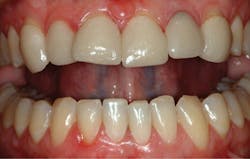
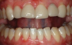
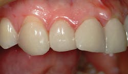
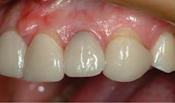
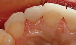
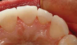
The following is an example of a patient who desired elective veneers to improve the appearance of her smile. She stated that her maxillary anterior teeth were spaced and rotated. Her lateral incisors were “pegged” and smaller than normal. The previous dentist was well meaning and did not prepare the teeth at all before taking a final impression for fabrication of porcelain veneers. In Figure 1, you can see the result that was obtained with six no-preparation veneers from canine to canine. Though the spacing was closed, the teeth still appeared malaligned and the shade was monochromatic. The close-up view in Figure 2 shows thick margins, some short of the tissue line. In Figure 3, you can see where there had been marginal leakage of the veneer on the lateral incisor evidenced by the black color. The incisal view in Figure 4 shows overcontouring of proximal areas and less-than-ideal tooth-veneer interfaces. Overlapping of proximal surfaces can be seen in Figure 5.
Figure 1: Six no-preparation veneers placed by the patient’s previous dentist
Figure 2: Margins in the previous restoration
Figure 3: Marginal leakage of the veneer in the previous restoration
Figure 4: Overcontouring of proximal areas
Figure 5: Overlapping of proximal surfaces in the previous restoration
Retreatment
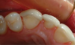
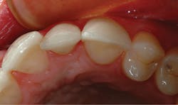
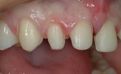
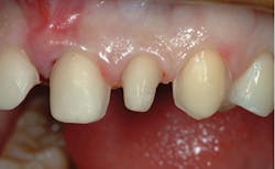
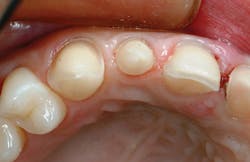
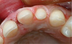
The previous veneers were removed by using a fine-grit tapered diamond bur in a high-speed handpiece, using copious water spray to keep from heating the porcelain. Care was taken to keep from removing any more tooth structure than was necessary to remove the veneers. A chamfer-ended diamond bur was used to define chamfer margins at the crest of the tissue. An incisal preparation allowing for at least a millimeter of overlap was accomplished with the same tapered bur. Proximal margins were ended at the lingual line angles to allow for wrapping of porcelain into the interproximal areas. The right central incisor was prepared with a 360-degree design to allow for better alignment. The left lateral incisor was also prepared for a 360-degree laminate. Figure 6 shows the right lateral view after preparation and in Figure 7 the left lateral view can be seen. The incisal views of the prepared teeth can be seen in Figures 8a and 8b.
Figure 6: The right lateral view after preparation
Figure 7: The left lateral view after preparation
Figures 8a and 8b: Incisal views of the prepared teeth
By allowing room for proper alignment and precise margins, porcelain veneers that mimicked a more natural appearance could be made. Ceramic veneers were fabricated for all six teeth and were bonded to place using a resin luting agent.
Practice management considerations: When a patient is unhappy with another dentist’s work
Debra Engelhardt-Nash
In the case described, the patient was dissatisfied with the results of her existing restorations and was seeking alternative treatment to be happy with her smile once again. How do you approach a patient who is searching for a better result than what was achieved with a previous dentist?
Instilling confidence in this patient begins the moment she calls the office. The telephone introduction should welcome the patient to the practice, instead of barraging her with questions. Asking about insurance coverage and launching into the rules and regulations of scheduling does not set the office apart as hospitable, nor does it make the patient feel as though she has called an exceptional practice.
Listen to the patient first; explain your office protocols second. What is most important during the phone call is what the patient wants to tell you, not what you tell her. Match her specific concerns or treatment interests with how your office meets those needs. Spend as much time as the patient needs to feel as though she has been heard and understood. This is not a timed event. Appeal to the human side of doing business with the patient, and your office standards will be more inviting.
The treatment consultation is a critical element to patient satisfaction and practice success. It is not an appointment that can be abbreviated or “sandwiched in” between other procedures. Whether a treatment coordinator or the doctor initiates this visit, proper time must be allocated to be sure the patient is comfortable and confident she has chosen the right office for her care.
Avoid denigrating the previous dentist and treatment. This does not elevate the status of your practice and creates a negative atmosphere for the patient. Explain your recommendations and focus on the expected results of your care. Tearing someone down does not build you up. Be the “bearer of good news” and show the patient her future potential. It’s a more winning approach.
Many people are visual learners. Present before-and-after images of actual cases done in your practice and show cases that are similar to the patient’s treatment. Help her visualize how she will feel with a new dental appearance. Spend the time with the patient to give her the confidence she needs to choose the care you both want.
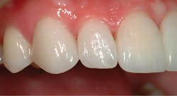
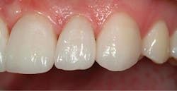
In Figure 9, the right lateral view of the final result can be seen. Figure 10 shows the left lateral view of the finished veneers. In the incisal view in Figure 11, the more natural contours of the porcelain laminates that have a better veneer-to-tooth juncture can be seen. In the incisal view in Figure 12, the 360-degree laminate on the left central incisor and the natural alignment of the anterior teeth can be visualized.
Figure 9: The right lateral view of the finished restoration
Figure 10: The left lateral view of the finished restoration
Figure 11: The incisal view of the finished restoration
Figure 12: The left central incisor view of the finished restoration
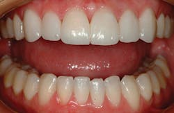
Figure 13: The finished restoration
Conclusion
In Figures 12 and 13, you can see that a natural color and translucency, excellent tissue health, and proper alignment of the six maxillary anterior teeth have been achieved. By preparing the teeth properly, the patient’s desire for a beautiful smile with well-aligned and natural-looking teeth was met. Sometimes we can be too conservative, compromising the end result. Even though some preparation was needed here, the amount of tooth structure removal was still kept to a minimum.
Ross W. Nash, DDS, maintains a private practice in Huntersville, North Carolina, where he focuses on esthetic and cosmetic dental treatment. He is an accredited fellow in the American Academy of Cosmetic Dentistry. Dr. Nash lectures internationally on subjects in esthetic dentistry and has authored chapters in two dental textbooks. He is cofounder of the Nash Institute for Dental Learning in Huntersville, North Carolina.
Debra Engelhardt-Nash is a trainer, author, presenter, and consultant, and has presented workshops, nationally and internationally, for numerous study groups and organizations. She is currently the vice-president and president elect for the National Academy of Dental Management Consultants, where she is a founding member and has served as president in the past. She was a contributing editor for Contemporary Esthetics and Restorative Practice magazine and Contemporary Dental Assistant magazine, and she has written for a number of dental publications.
