Shade Matching for Today's Dentistry
The science and art of color perception, interpretation, and reproduction
by Jean Sagara
What is the problem? Ask yourself this question: How many times has a patient left my office after a cosmetic treatment and I said to myself, "That could have been better"? Too many times, we dare to suggest. The reason is simple enough. While we practice the science of dental medicine, increasingly we find ourselves promising to deliver art — the art of the beautifully transformed countenance. We sell smiles.
Selling smiles that achieve full patient satisfaction can be challenging. Success or failure largely depends on the accurate transfer of subtle information from the dentist to the laboratory. Sometimes this information exchange goes poorly. Dental laboratories admit to a 6 percent remake rate annually as a cost of doing business. Half of these remakes (3 percent) occur for two reasons: 1) as a result of misinterpretation of critical data, or 2) as a result of a failure to match shades accurately.
For the dentist, misinterpreted data can be overcome with full and careful communication with the lab. The failure to match a patient's tooth shade, however, is not as easily fixed.
Color and shade are subjective values. When they aren't right, it is quite obvious and very personal. What the patient eagerly anticipated as a natural-looking enhancement is instead an unflattering clinical failure for the professional.
A: A little too often.
Shade: An elusive attribute
Shade matching involves many variables, some of which are not easily controlled. It also involves a delicate balance between managing realistic goals against personally held expectations. The stakes are high and mistakes can be costly to the patient, the dentist, and the lab. Consumers perceive an attractive smile as a necessary part of a confident presentation of themselves. The most readily observable part of the body is the face. Enhanced by a beautiful smile, people tend to feel good about themselves. When the shade match is wrong, with variations in color or opacities and myriad other details, it can give a person's smile the look of "missing the mark." The eye can detect these discrepancies. Successful shade matching is science and art combined. It involves understanding the science of color, determining surface characteristics, and communicating this subjective information so that it is objectively understood by the laboratory technician. It also involves a partnership with your patient to set reasonable goals.
Shade can elude exactness because it has a layered quality. Fundamentally, there are three elements to a shade: Hue is the "color;" chroma is the concentration or saturation of a hue (intensity); and value is the lightness or darkness of a hue, usually measured in variations of gray. In our many conversations with colleagues, it becomes clear that value often can be the most important. Dean Ribeiro, group manager for National Dentex Corporation, the country's largest dental laboratory organization with 28 labs located throughout the United States, said, "Of all the remakes due to improper shade selection, we find that 70-75 percent fail because the value is off." In fact, to address this issue, National Dentex offers a course called "Getting on the Same Wavelength" to dentists and dental students. It offers practical guidance on how to optimize shade selection, including simple tips guaranteed to minimize the risk of remakes.
How is shade matching done today?
Shade matching is a matter of color perception and subjective interpretation. For example, make the color "bluer" or make it "greener." But, bluer or greener than what? That's the question. The prosthesis usually is created away from the patient's personal attributes of hue, chroma, and value. Thus, capturing that individual's shade is challenging. Progress is occurring, however, and our tools are getting better and better.
Let's review some standard procedures for obtaining a shade, and look at some of the new techniques. Detailed information on these and other resources is available at www.transcendonline.com.
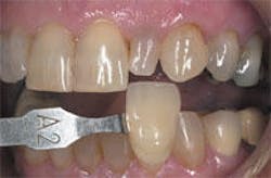
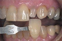
Shade tabs. In the past, shade matching was a combination of impressions, shade tabs, and a written prescription. The dentist held the shade tab next to the patient's teeth, wrote its number on a piece of paper, and passed it along to the technician for a "perfect match." This was an oversimplified approach that almost guaranteed an aesthetic failure, because too much was left to subjective interpretation and too little was offered in terms of a real standard — a baseline from which to begin. While it is true that most shade guides offer a color order arranged by hue groups and divided by chroma levels, the most useful color dimension — value — often is not considered. And as most dentists will attest, a "perfect match" is a combination of several shade tabs, not just one.
Shade guides are improving, however. Vitapan 3D-Master Shade Guide presents a new variation on the shade tab system. This guide presents almost complete coverage of the tooth color space and gives the dentist three steps as opposed to the traditional one step. Here is how it works. First, the dentist determines the value (dark/light) from among five value groups; then determines the chroma (color saturation/intensity) from among three chroma groups; and, lastly, determines the hue (color); that is, is there more yellow or red cast in this tooth? Shade guides with this degree of differentiation begin to give us a reference point, but they are not the whole answer. As George Zoller, CDT, of Anderson Dental Labs, observes: "Teeth are wonderful mosaics of color. We must use the shade guide as one piece of the puzzle. They map to patterns we see ... they are a stable reference point and begin the process." Recognizing the difficulty associated with shade matching, Americus Dental Laboratory, the nation's third largest dental lab, gives a course on color mapping to support its customers and maximize quality outcomes.
Photography and imaging. Photographs are helpful to the dentist and the lab in communicating key shade information. Bob Moyer, president of Stern Reed Labs, a National Dentex Reliance Qualified Laboratory located in Dallas, Tex., admits that "... clinical photographs are very helpful with characterizations and texture ... even though the actual shade is not perfect in the photo." Practically speaking, photographs are essential for anterior teeth detail on size, shape, translucency, and other individual characteristics. Many dentists still rely on 35 mm photography, using professional-quality equipment such as a single lens reflex (SLR) camera and film such as Kodak photographic print or slide film. This film is manufactured to ensure consistent color and image quality. When used in conjunction with a professional color lab for photo processing and a camera that produces high-resolution images, these same images, when sent to the lab, can reveal additional information critical to a successful outcome.
Shade tabs and photographs are, of course, very complementary. To record an objective color on film as closely as possible, a shade tab is placed near the patient's mouth and photographed from a frontal view. This image then can be used as a baseline for the technician who can hold the same shade guide next to the print and adjust the shade of the actual crown, for example, according to any distortions that appear.
Dr. Edward B. Walk, one of the industry's most respected experts on the use of photography in dentistry, offers appropriate cautions about the limits of images, however. He told us, "... the problem is usually that what you see is not necessarily what you get." By this, he means that the visual image of the actual tooth is not precisely the same as the photographic image of that same tooth. In other words, the eye beholds something different from what the lens can capture. Still, it is readily acknowledged that images go a long way toward improving the quality of the information exchanged between a dentist and the lab. Dr. Walk has perfected a photo-layering technique on which he lectures nationally, and which he believes minimizes the obvious limitations of the art form. Visit www.transcendonline.com for more information on his specific technique.
Digital imaging. While instant photographs, and 35 mm slides and prints are still used, digital photography is inserting itself more and more into the practicing dentist's armamentarium. Using a digital camera with a charged coupling device (CCD), the dentist can capture images directly onto a personal computer. From there, images can be saved, uploaded for sharing with the lab online, and archived as a part of the patient's personal file.
For example, Americus Dental Lab's eLabRx, an online prescription service that applies smart-form technology to prescription work flows, capitalizes on image information. Using image uploads with intuitive Web designs, complete instructions go to the lab for easy scripting in real time.
Sometimes digital images can reveal more clinical data than a standard 35 mm photograph. Of course, the greater the resolution of the camera, the better the image provided. This applies to all forms of input devices. In the end, digitizing information, particularly images, gives the dentist some secondary benefits. With images stored on a local directory, the dentist can use them in referrals or for ongoing treatment planning. They also can come in handy when dentists need to prepare lectures for seminars with their colleagues.
Increasingly, dentists are appreciating the value of images to enhance the information exchanged with their laboratories. Dr. David Thibault, a general practitioner from Greenwich, Conn., remarked that "... including clinical images makes dentistry much more predictable for me and increases my confidence as well as that of my patients."
Dr. Phil Morisseau, another general dentist from North Kingston, R.I., agreed. He offered, "I include images with my lab cases because the more information I give the lab, the better the result."
The dental labs interviewed for this article concurred. Art Francis, sales and marketing manager for Arcari Dental Laboratory, is very clear on the contribution photographs make toward an excellent result. "We receive color photos as well as tooth casts and shade selections. Incoming images are automatically presented to the technician. Comments can be added to the image and returned to the dentist for clarification if needed. Photos from film or instant cameras, digital cameras, and intraoral cameras can be used with ease."
How can we measure results?
Where does that leave us? The labs tell us that half of the remake rate or 3 percent is due to mistakes with data interpretation and shade mismatching. The lab and the dentist bear the costs. The color industry tells us that 100 percent color matching is impossible. Dentists tell us that in addition to the 6 percent total remake rate, the outcomes for patients vary from "That's very nice" to "It's good enough." But is "good" really good enough?
Dentists need to persist with simple, sustained commitments as their labs also look for ways to improve customer service. Professionals should continue to learn color-mapping techniques, increase their photography skills, and improve their overall lab exchanges with enhanced image and information communications. Technology investments must not be sidelined. We know that more dentists are purchasing digital cameras, spending anywhere from $400 to $6,000. Some of the color-mapping devices and software products currently on the market require an investment of between $4,000 and $15,000, with dentists as well as laboratories taking the plunge. At the other side of the equal sign is the return on investment, which is a personal calculation. To consider ROI, you must ask three questions: 1) Does the technology improve the outcome? 2) If so, at what cost? 3) Are there measurable benefits?
As we move toward higher standards with shade-matching techniques, Lee Culp from the Institute of Oral Art and Design reminds us that certain fundamentals remain. "The future holds great promise with computer-based shade systems that allow for more objective measurement of color, value, and translucency without the inaccuracies of environment or human color perception. With that said, these systems will not overcome an inaccurate preparation done by a dentist, or assist a ceramist who does not have the training or understanding of his materials to create a natural-looking restoration. A complete understanding of color, materials, and tooth anatomy is essential for creating natural-looking restorative dentistry."
In the end, it turns out that dentists practice both science and art. Advances in one aspect bring greater mastery over the other. This has always been our truth, but its constancy is gaining as more and more consumers are becoming our patients, looking for that pleasing countenance.
For more information on shading systems, be sure to check out the January 2002 issue of Dental Equipment & Materials, a sister publication of Dental Economics.
Emerging technologies
Public awareness of the possibilities associated with restorative dentistry has led to the expectation that people with prosthetic teeth and restorations can function and look better than they do with their own teeth. Many of these expectations come from the development of materials and technologies. Central for dentists is still the question: How do we give cosmetically minded patients greater confidence in their restorations? One way is through continuing education and attention to emerging technologies. The dental professional is constantly faced with buying decisions for products and technology. In some cases, the decision is easy. If the product provides the ability to do something that previously was not possible, or solves a problem while also providing a new source of income, the decision is easy. If the dentist can identify a product that improves outcomes and increases incomes, the choice seems obvious. What could be better than to elevate standards, patient satisfaction, and revenues all at the same time?
With shade matching in particular, there are some new technologies on the horizon. The patented SpectroShade System from MHT International allows for complete aesthetic information to be communicated from the dental practice to the laboratory. It uses a spectrophotometer to deliver scientifically accurate shade measurements (6 million data points per image), clearly showing the variance between the closest shade reference and the actual tooth shade. Another system called the ShadeScan System by Cynovad, Inc., is a hand-held image-capture device and vision software system designed to do color mapping to produce an optimal visual appearance of patients' teeth. Another product called the Shade Vision System by X-Rite, Inc., the world's largest manufacturer of industrial color-measurement devices and just entering the dental market, eliminates the variability of human color perception and advances color difference communication. As Tom Nyenhuis, vice president of sales and new business development at X-Rite, Inc., observed, "Once dentists and labs begin to measure and communicate in the language of color (hue, chroma, and value), the process improves and the result is a better product. Instrumentation is the key." (www.transcendonline.com has more information on these and other systems.)

