Pinhole Surgical Technique: A 10-year evaluation of root coverage
Gingival recession is a soft-tissue problem affecting 88% of adults in the United States aged 65 and older and 50% of adults aged 18–64.1 Gingival recession is defined as the displacement of the marginal tissue apical to the cementoenamel junction (CEJ), where the amount of tissue that is lost (apically displaced) between the CEJ and the gingival margin is the amount of recession present.2
Causes of gingival recession include toothbrush and oral hygiene abrasion, periodontal tissue loss, calculus, high frenum attachments, position of the tooth in the arch, trauma, orthodontic movement, smoking and tobacco products, genetically thin tissue, subgingival restorations, poorly fitting dentures, and chemical abrasion from medications/drug use.3
Also by the author:
- Dental implant contamination: 3 reasons behind a late-stage failure
- Smile makeover: A minimally invasive recipe for esthetic success
Consequences of untreated tissue recession include poor esthetics (long-looking teeth), root hypersensitivity, root caries, plaque retention, bleeding, and continued tissue loss.4
Various soft-tissue grafting surgical techniques to treat gingival recession exist, including free gingival grafts, connective tissue grafts, pedicle grafts, rotational grafts, coronally advanced flaps, guided tissue regeneration, and other modifications to these standard grafts.5 The disadvantages of many of these techniques are they are invasive, may not result in perfect tissue replacement color-match, or may require a second donor site to harvest the graft tissue.6
Pinhole Surgical Technique
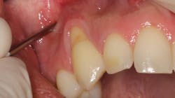
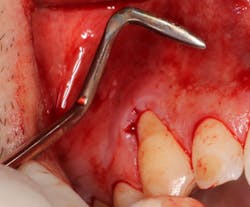
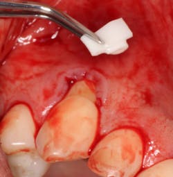
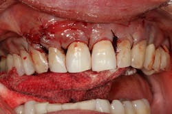
Introduced by Dr. John Chao in 2012, the PST is a minimally invasive soft-tissue grafting approach using microincisions (pinholes) in the vestibular tissue (figure 1). Specialized transmucosal periosteal elevators (figure 2) are then used to advance the existing soft tissue coronally to cover the recession defects. Graft material, typically porcine collagen membrane (Geistlich Bio-Gide), is insertedResearch about PST
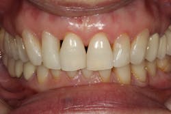
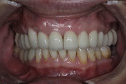
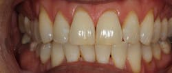
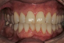
In 2012, Dr. John Chao, the inventor of PST, published the first study using this technique on 121 recession defects. He noted that for teeth with early and moderate recession with no bone loss (class I and class II Miller recession defects), recession was reduced about 94% and complete root coverage wasConclusion
The PST has been shown to have excellent results over a long-term period and is comparable to other soft-tissue grafting techniques. Advantages include aAuthor’s note: Dr. Scott Froum is not a paid consultant and does not have any financial interest in any company mentioned in this article.
Editor's note: This article appeared in the May 2022 print edition of Dental Economics magazine. Dentists in North America are eligible for a complimentary print subscription. Sign up here.
References
- Miller AJ, Brunelle JA, Carlos JP, Brown LJ, Löe H. Oral health of United States adults. The National Survey of Oral Health in U.S. Employed Adults and Seniors: 1985-1986, NIH publication no. 87-2868.
- Glossary of Periodontal Terms. AAP Connect. American Academy of Periodontology. https://members.perio.org/libraries/glossary
- Koppolu P, Rajababu P, Satyanarayana D, Sagar V. Gingival recession: review and strategies in treatment of recession. Case Rep Dent. 2012;2012:563421. doi:10.1155/2012/563421
- Chambrone L, Tatakis DN. Long-term outcomes of untreated buccal gingival recessions: a systematic review and meta-analysis. J Periodontol. 2016;87(7):796-808. doi:10.1902/jop.2016.150625
- Goyal L, Gupta ND, Gupta N, Chawla K. Free gingival graft as a single step procedure for treatment of mandibular Miller class I and II recession defects. World J Plast Surg. 2019;8(1):12-17. doi:10.29252/wjps.8.1.12
- Camargo PM, Melnick PR, Kenney EB. The use of free gingival grafts for aesthetic purposes. Periodontol 2000. 2001;27(1):72-96. doi:1034/j.1600-0757.2001.027001072.x
- Zadeh HH. Minimally invasive treatment of maxillary anterior gingival recession defects by vestibular incision subperiosteal tunnel access and platelet-derived growth factor BB. Int J Periodontics Restorative Dent. 2011;31(6):653-660.
- Griffin TJ, Cheung WS, Zavras AI, Damoulis PD. Postoperative complications following gingival augmentation procedures. J Periodontol. 2006;77(12):2070-2079. doi:1902/jop.2006.050296
- Reddy SSP. Pinhole Surgical Technique for treatment of marginal tissue recession: a case series. J Indian Soc Periodontol. 2017;21(6):507-511. doi:10.4103/jisp.jisp_138_17
- Agarwal MC, Kumar G, Manjunath RGS, Karthikeyan SSS, Gummaluri SS. Pinhole Surgical Technique — a novel minimally invasive approach for treatment of multiple gingival recession defects: a case series. Contemp Clin Dent. 2020;11(1):97-100. doi:10.4103/ccd.ccd_449_19
- Mostafa D, Al Shateb S, Thobaiti B, et al. The pinhole technique in the treatment of gingival recession defects. A long-term study of case series for 5.1–19.3 years. Adv Dent Oral Health. 2020;13(1):555855. doi:10.19080/ADOH.2020.13.555855
About the Author

Scott Froum, DDS
Scott Froum, DDS, a graduate of the State University of New York, Stony Brook School of Dental Medicine (SUNY), is a periodontist in private practice in New York City. He is the editorial director of Perio-Implant Advisory and serves on the Dental Economics advisory board. Dr. Froum is a volunteer professor in the postgraduate periodontal program at SUNY and a PhD candidate in the field of functional and integrative nutrition. Contact him at drscottfroum.com.
Read Dr. Froum's DE Editorial Advisory Board profile here.
