Arturo R. Garcia, DMD
The microscope is the most important, productive, and frequently-used piece of technology I have. All technologies and techniques have an indicated application; that is, they have a specific use. The microscope is unique in that it is the one piece of technology that can make all others technologies and techniques better. Why? Because no matter what procedure you are doing, you need great visualization to do it. The dental microscope is the best way to achieve great visualization; the better visualization you have, the better results you get. I also do all of my endo and perio treatments under the scope.
I have been using a microscope for three years. I first started using magnification in 1990 when I bought a bench microscope to use in my dental lab. It was a 10x microscope and I used it to examine the crown dies that came from my impressions. Wow! I was amazed at the difference between what I saw clinically without the aid of any magnification and what I saw under the bench microscope at 10x. I could not believe what my preps looked like under the microscope relative to what I remembered seeing and doing clinically without any magnification. To say the least, I was horrified!
In 1992, I started using 3.5x loupes clinically and was amazed at the difference they made. My clinical awareness went way up! I could not believe what I had been missing without them. I wanted more magnification and got a pair of 4.5x loupes. This was a big improvement, but the more I used them, the more I realized how they were getting heavy and cumbersome. At this level of magnification, good lighting is critical and difficult to achieve with the standard patient light. However, the benefits of the increased magnification were awesome. My level of clinical awareness went up again. I was amazed at what I had been unable to see before with lower magnification.
Seven years of experience with the benefits of loupes left me wanting to use higher magnification. I noticed that high quality labs routinely used bench microscopes to produce the high quality restoration that I requested. Endodontists routinely and successfully used microscopes to improve their treatment results. This resulted in a quantum leap forward for endodontics. I briefly considered 7x loupes, but was unwilling to put up with a heavier, more cumbersome set. I also was concerned about adequately lighting the treatment field. I already was struggling with the 4.5x loupes and knew good lighting would be even more problematic at 7x.
Finally, I did not want to be confined to one power of magnification, which would not serve all of my clinical needs. I wanted the ability to observe and evaluate at low power. Then, a higher power could be used for working and inspection. I wanted to do all of this in a well-illuminated field and be able to reach any area of the mouth comfortably.
Aesthetic/restorative dentists continuously strive to produce the best results possible for our patients. Perhaps the most important part of producing the best results is getting the best information that you can about what you are doing. A thorough, pretreatment patient examination is essential. The visual part of this exam is an important, yet often underutilized, part of the examination process. I use the word "underutilized" because there is so much more information available than what meets the eye. You certainly get information when you examine your patient without magnification, but think about how much more information you could get using magnification. Even at low power magnification (2x loupes), you are getting 100 percent more detail about what you are seeing compared to no magnification. More information results in a better product!
Many offices now use intraoral cameras for their visual examinations. Why? Because of the great visualization (high-power magnification and great lighting) that intraoral cameras provide. This is far superior to the naked eye.The intraoral camera's great visualization allows us to get much more accurate information and to educate our patients with clear pictures. We go to great lengths to collect as much accurate information as possible - i.e., great color images from the intraoral camera, high quality digital or analog X-ray images, and great study models mounted on the Acculiner with a comfortable bite. Tremendous effort goes into collecting accurate and high quality information about our patients. From this information, we formulate a comprehensive treatment plan.
If the dentist is not using magnification during treatment, the benefit of the quality of information that was collected at higher resolution is diminished or lost. If an intraoral camera was used to collect color images - and treatment decisions were made from these high magnification images - we need to carry that level of awareness through to the actual treatment. Great visualization is important for diagnosis, but it is critical for treatment.
We cannot diagnose or treat that which is below our level of awareness. One reason we use modern technology is to elevate our level of awareness and bring the level of treatment up to that of the disease we are treating. Improving our ability to see - or improving our visualization - is central to elevating our level of clinical awareness. Once dentists embark on the path of improved visualization, starting with loupes, they won't go back to working without enhanced visualization.
What is great visualization?
Great visualization is the product of high magnification combined with high-intensity lighting. The high-intensity lighting has to be coaxial; that is, the direction of the light is parallel to the direction of the line of sight. Coaxial lighting is important because as the level of magnification is increased, the field of view becomes darker. Loupe users usually start out at low power (2x). As they go to higher-power loupes, they start to implement high-intensity light sources to compensate for the darker field of view at higher magnification. These light sources are either clipped on the loupe frame or on a headband. This combination of loupes and light source becomes cumbersome and restrict movement.
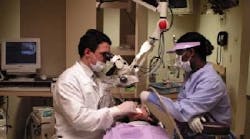
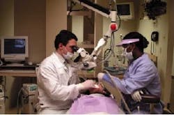
When dentists use loupes consistently (that is, they use their loupes all the time, not just for inspection) and want to go above 3x in power, they are ready to move up to a dental microscope. One of the great attributes of the dental microscope is that it has multiple levels of magnification (five steps) and a built-in coaxial, high-intensity light source. No matter where in the treatment field you look, you always have great lighting without the need to use a patient light. In fact, in my office, I don't have overhead patient lights! I use my microscope for almost everything I do, so I don't have a need for patient lights. When I'm not using the microscope for visualization, I use it as a patient light.
The use of microscopes in an aesthetic practice
The key to successful integration of the dental microscope into an aesthetic/restorative practice is training. Using a microscope is much different than using loupes. The dentist and the assistant need to be trained on how to effectively and efficiently use a microscope. Experienced microscope users estimate that a clinically relevant microscope course (a course that has a clinical component - not just a bench top or over-the-shoulder course) can shorten the new microscope user's learning curve by 6-12 months.
A course of this type is available at the Las Vegas Institute. It is a three-day, live patient course, designed to train the dentist and his assistant. Most new, untrained microscope users become very frustrated with using the microscope because they don't understand the fundamentals and tend to give up easily. It's like trying to do a veneer case without understanding the principles of bonding or smile design - not too much fun!
Most experienced microscope users comment with amazement about the wonder of working through a microscope. In my practice, it has rejuvenated my commitment to quality dentistry. The dental microscope is also the one piece of technology that my patients understand and identify most easily. Most people have either seen or used microscopes during their education or have seen them used in medicine. That makes most patients very accepting and understanding of the commitment to excellence embodied by using a dental microscope.
Benefits of visualization
The following benefits are the result of using any type of visual enhancement. However, these benefits are more typical of a dental microscope than any other type of visual enhancement, due to the superior visualization provided by the microscope.
Improved efficiency - The great visualization provided by the microscope results in an elevated level of awareness at the diagnostic level and an elevated level of efficiency at the treatment level. The increased awareness at the diagnostic level is much like the benefits of an intraoral camera. The increased efficiency at the treatment level is the product of great visualization. You can comfortably reach and see any area of the mouth. There is no question about what you set out to do - it is clear when you have accomplished what you intended.
Improved quality of treatment - More than 70 percent of the treatment that restorative dentists perform involves replacing existing restorations. The choice between direct or indirect restorations often depends on the amount of remaining tooth structure. Often, tooth structure looks sound at macroscopic levels. But, under high magnification, the it may become obvious that the remaining structure is full of micro fractures that extend from the internal surface of the preparation out to the finish line. These fracture lines, if not accounted for in prep design and restoration choice, often can lead to premature failure of the restoration, sensitivity, or recurrent decay. This is especially critical when using bonded or cemented restorations, because these fracture lines are water permeable and allow for the continued infiltration of bacteria.
Our medical brethren (ENTs, neurosurgeons, etc.) have been using high-powered microscopes for over 50 years. Microscope users receive much more visual information about the treatment area than without a microscope. This increased information results in the development of finer neuro-motor skills - i.e., a more delicate, deliberate, and precise touch. Studies have shown that magnification users develop finer neuro-motor skills and, as result, make fewer mistakes than those who don't use magnification.
Improved ergonomics - Back, neck, and shoulder damage are a leading source of physical disability for dentists. The majority of these disabilities are due to years of poor posture and positioning as dentists struggle to see what they are doing. When you see dentists who work without magnification, you notice that they typically work in a bent-over position. Their torsos are twisted, and the lower back and shoulders are strained as they struggle to see what they are doing. Dentists who wear loupes typically exhibit a more upright posture. They sit in a comfortable and upright position with little neck, back, or shoulder strain. This is due to the greater access and visualization provided by the microscope. This increased comfort also improves the patient experience; the dentists working with the aid of a microscope use micro movements and lighter pressure. Without a microscope, rougher macro movements are produced.
Increased productivity - When you can work in a physically comfortable position for you and your patient and you can easily reach and see what you are doing, you become much more productive. The microscope enables the dentist to work in a focused fashion with direct movements and fewer wasted movements.
Improved stamina - The increased comfort that comes from using the dental microscope allows the dentist to work comfortably and efficiently for extended periods of time. The dentist is physically comfortable due to the correct posture produced by using the dental microscope. The dentist also experiences little or no eye strain, due to the great visualization provided by the microscope.
