Increasing patient acceptance of smile makeovers with minimally invasive veneers
by David Silber, DMD
When evaluating the goals of a dental practice, one aspect that resurfaces often is improving patient acceptance of cosmetic treatments. After all, what patient does not want to improve his or her smile? Today, through media exposure, patients are better educated about smile improvement via teeth bleaching and veneers. As a practitioner, the goal of improving patient acceptance for cosmetic treatment may not meet your expectations because patients demand more.
Questions will arise when your expectations of treatment acceptance have not been met. One question might be, “Why doesn’t the patient accept the cosmetic treatment plan that is presented?”
Perhaps the presentation of the treatment plan by your staff and you is lacking. Perhaps your office environment does not communicate esthetic treatment well to the patient. Some practitioners seek expert advice from consultants and colleagues. They want that special tidbit of information - how to offer the perfect smile makeover solution. It would be a perfect world if there was just one concept, one presentation tool that would achieve that magical 100 percent acceptance of treatment plans presented.
While that ideal does not exist, there is magic in planning your case presentation to educate patients about the benefits of treatment, final outcome, and expectation of the proposed treatment. I have found that the more visual aids you provide the patient, the higher the percentage of cases that will be accepted. By using a patient in my practice as an example, I will demonstrate visual enhancement techniques in my office that have allowed my staff and me to turn a critical corner toward improved case acceptance.
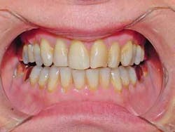
A female patient came to my office seeking whiter, brighter teeth (Fig. 1). Her experience with a previous dentist involved a painful one-hour whitening session that left residual sensitivity. The result of the whitening did not meet her expectations. Without any alternatives, she was willing to go through the whitening procedure again - even tolerate the pain she experienced the first time. I needed to understand, in detail, what brought her to my office since it was obvious to me that there was much to understand about her story.
Many times patients will not open up and let you know what they are thinking. For me, the initial encounter presents a challenge to break the ice with the patient and let the person know he or she can feel comfortable talking with you. A patient must know he or she can “spill the beans” about any problem(s). This type of patient understands there is no judgment concerning past dental experiences. Your goal is to help the person understand and achieve his or her desired goal. For this patient, and many others, the question is simple: “Do you like your smile?” This one question can open the floodgates.
For my patient, the key turned and the door opened when I asked “Do you like your smile?” She responded that she hated her smile, did not want to smile, and was embarrassed by her smile. She described how she wanted to be out of sight when anyone was taking a photograph. She offered a vivid description of what she wanted.
“I hated whitening because it hurt, and I am not interested in veneers because I know that it will involve drilling my teeth,” she said. “Is there anything that can be done to improve my smile without the pain?” My discussion about the experiences with her previous dentist and the pain associated with treatment made me aware that I needed to offer a painless solution to this overwhelming problem.
As part of my consultation and examination visits, I make a full-mouth series of radiographs, a complete series of digital clinical photographs, impressions for study casts, and a bite registration. We rescheduled the patient for a treatment plan presentation appointment. (As part of our office policy, her smile makeover evaluation was billed to her, but we explained that this fee would be applied to future treatment upon her acceptance of the treatment plan.) In my practice, the key to the smile makeover evaluation is to create an aid that helps educate our patients on the goals of final treatment. The digital photographs are used to create a preview of the final smile makeover, and to create a desire for the procedure. The study casts are used to create a diagnostic waxup, or a roadmap, for the smile makeover and provide a guide for any tooth modifications.
In some cases, a template for provisional restorations for more involved prosthodontic treatment is necessary. For our example (and from my past experiences), I knew that minimally invasive veneers were the best option to meet the patient’s expectations of a painless, permanent solution for her smile makeover. Minimally invasive veneers provide a stronger, more predictable bond because they are bonded directly to enamel. This also results in no postprocedure sensitivity. I also knew that, for this patient, seeing what could be done to change her smile would result in treatment acceptance.
Where do you get the expertise for creating a digital image and study cast preview for the final case presentation of a patient’s smile makeover? My experience involving digital imaging services with my laboratory (Cerinate Smile Design Studios) has been excellent. The laboratory specialists have the experience and knowledge to assist a clinician in planning cases. In this case, I sent the casts to my laboratory for consultation, evaluation, and a diagnostic waxup (based upon my directions) noting what the patient wanted to achieve as a final smile outcome.
Through the waxup process, the laboratory made recommendations for tooth modifications. While the smile design diagnostic waxup helps a patient visualize his or her new smile, my experience has been that digital photographs demonstrating the transition from current smile to new smile make the biggest difference. As part of the process, I e-mail the digital intraoral photographs to my lab. Based upon the esthetic diagnosis with a patient, the smile preview is digitally imaged and then returned to me via e-mail. For this patient, the use of both teaching aids was critical to the success of the case presentation and case acceptance.
Once I had all the support materials from the laboratory, a digital image showing before-and-after photos and the diagnostic waxup, I presented a treatment plan of some minor soft-tissue reshaping and the placement of minimally invasive veneers to achieve the smile she desired.
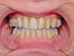
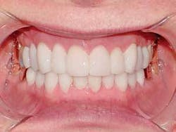
Per my request, the digital images included one preview with no gingival changes (Fig. 2) and one preview with minor gingival alteration (Fig. 3). It is important for a patient to understand the cosmetic benefits for each of the previews and treatments involved.
With the aids provided, a patient is better able to visualize his or her case and the benefits of the minor soft-tissue procedure when combined with minimal tooth reshaping for the placement of the minimally invasive LUMINEERS. In our example, at the end of the treatment presentation, the patient accepted the gingival tissue recontouring along with the placement of the noninvasive veneers. She chose the shade for her new teeth, and appointments were set.
After the minor gingival recontouring was performed and had ample healing time, this patient was ready for the placement of the minimally invasive veneers. Following the lab’s recommendation, the teeth were reshaped in a painless procedure without the need for local anesthetic. Then a full-mouth impression was made. This revealed sharp, easy-to-read gingival margins. The beauty of no-prep veneers is that a patient can leave the office without the need for temporary restorations. Patients appreciate the benefits of conserving their natural tooth structure and ability to forego the use of temporaries that can break while waiting for the final case. The impression was sent to the lab, along with the digital images and the esthetic diagnostic waxup intended to serve as a guide for the laboratory artists.
My instructions to the laboratory included the use of Cerinate Pressed Porcelain because of its excellent strength and long-term clinical success, durability and matched physical properties with the tooth and bonding materials.
The minimally invasive veneers were custom-fabricated and returned. Then they were tried in for final patient acceptance. The patient was excited and could not wait for the final result. When the veneers were placed, and the patient saw the final result, she was ecstatic (Fig. 4).
Minimally invasive veneers are my choice for veneers. Research indicates that they can last more than 20 years with porcelain that has physical properties matched to tooth structure and bonding materials. I have confidence in the long-lasting smiles that these no-prep veneers can create.
Increasing patient acceptance of esthetic treatment cases is a goal of any practice. With the use of aids, such as digital imaging and esthetic diagnostic waxups, patients can visually enhance their understanding of treatment outcomes for a smile makeover. Through the use of visual and tactile aids and minimally invasive veneers, dentists also can see an increase in patient acceptance of esthetic treatment since the pain factor is removed while the visual desire is created.References available upon request.
David Silber received a DMD degree from the University of Puerto Rico in 1994. He lectures for the Den-Mat Corporation. His lecture subjects include cosmetic dentistry, air abrasion techniques, and dental office management. Contact him by e-mail at [email protected] or via the Web at www.planodentaloffice.com.


